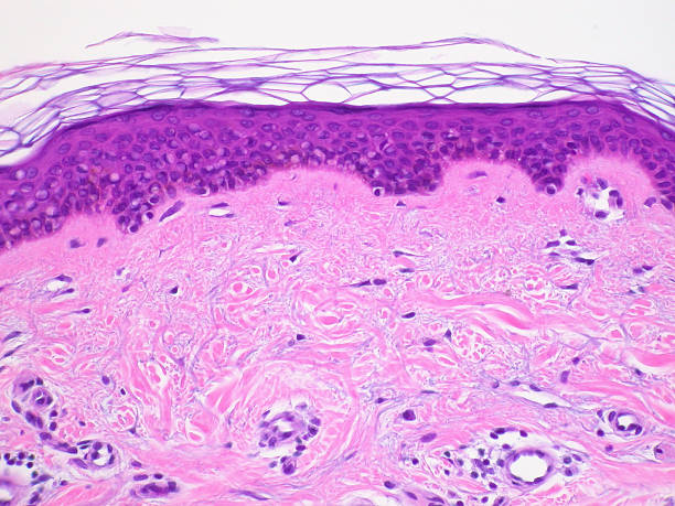
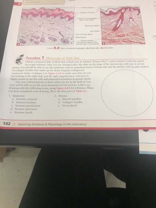
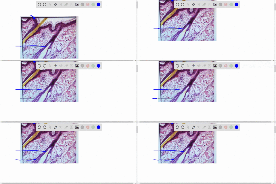
41 photomicrograph of skin labeled
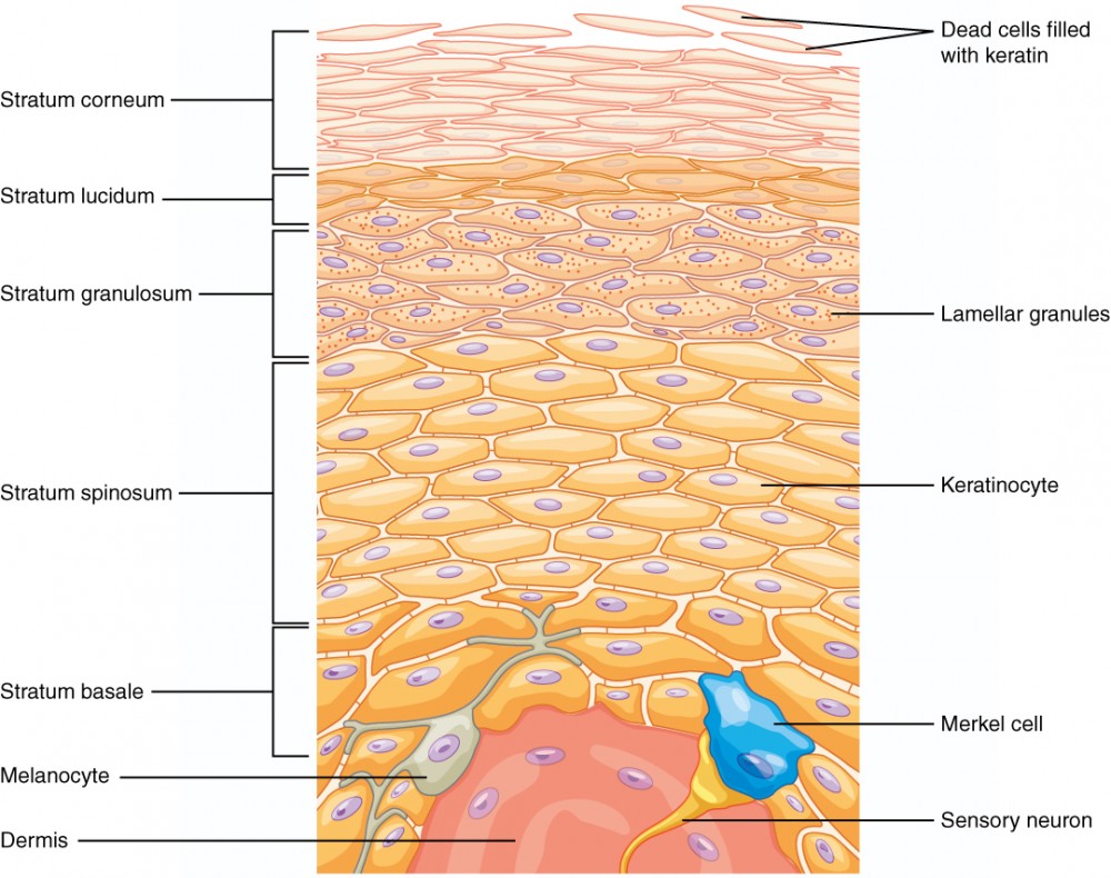
Solved Label the photomicrograph of thick skin | Chegg.com Anatomy and Physiology questions and answers Label the photomicrograph of thick skin Question: Label the photomicrograph of thick skin This problem has been solved! See the answer Show transcribed image text Expert Answer 91% (11 ratings) Transcribed image text: Label the photomicrograph of thick skin Previous question Next question 5.1 Layers of the Skin - Anatomy & Physiology Skin that has four layers of cells is referred to as "thin skin.". From deep to superficial, these layers are the stratum basale, stratum spinosum, stratum granulosum, and stratum corneum. Most of the skin can be classified as thin skin. "Thick skin" is found only on the palms of the hands and the soles of the feet.
Label The Photomicrograph Of Thin Skin. - Skin Model 1 - YouTube Label the photomicrograph of thin skin 1 answer below ». The ducts are lined by stratified (2 layers) cuboidal epithelium. Hair sebaceous gland dermis hair follicle epidermis duct of sebaceous. Label the photomicrograph of thin skin. This is a photomicrograph of thin skin. Learn more about thin skin treatment at howstuffworks.
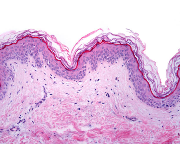
Photomicrograph of skin labeled
Figure 7.1: Photomicrograph of Skin Diagram | Quizlet Figure 7.1: Photomicrograph of Skin 3.0 (2 reviews) + − Learn Test Match Created by SophiaVisaggio Terms in this set (5) dermal papillae ... epidermis ... papillary layer of dermis ... reticular layer of dermis ... hypodermis ... Sets found in the same folder photomicrograph of the epidermal layer i… 6 terms abba_dabba_17 Diagram of human skin structure — Science Learning Hub Diagram of human skin structure. Image. Add to collection. Tweet. Rights: University of Waikato Published 1 February 2011 Size: 100 KB Referencing Hub media. The epidermis is a tough coating formed from overlapping layers of dead skin cells. photomicrograph of thick skin Diagram | Quizlet photomicrograph of thick skin Diagram | Quizlet photomicrograph of thick skin + − Learn Test Match Created by mckennawebber Terms in this set (7) epidermis (stratum corneum - stratum basale) ... stratum corneum ... stratum lucidum ... stratum granulosum ... stratum spinosum ... stratum basale ... dermis ...
Photomicrograph of skin labeled. Label The Photomicrograph Of Thick Skin - Lichen 4 The Algal Layer ... This is a picture of an h&e stained section of the epidermis of thick skin. 1 answer to label the photomicrograph of thin skin. Cornified (keratinized) stratified squamous epithelium makes up the epidermis. It has a fifth layer, called the stratum lucidum, located between the stratum corneum and the stratum granulosum (figure 2). Layers of the Skin | Anatomy and Physiology I - Lumen Learning Skin that has four layers of cells is referred to as "thin skin.". From deep to superficial, these layers are the stratum basale, stratum spinosum, stratum granulosum, and stratum corneum. Most of the skin can be classified as thin skin. "Thick skin" is found only on the palms of the hands and the soles of the feet. (Solved) - Label the photomicrograph of thin skin. Label the ... Label the photomicrograph of the skin A photograph taken with the help of microscope . Skin is the largest sensory organ in body.its protect the body sense pain sense temperature and pressure. STRUCTURE OF THE SKIN Epidermis-outer most layer of dead skin cells,prevents the body from losing water protect the body against infections. Photomicrograph of Thick Skin Quiz - PurposeGames.com Photomicrograph of Thick Skin Quiz Science » Image Quiz Photomicrograph of Thick Skin by nhammond21 1,701 plays 6 questions ~ 20 sec 2 too few (you: not rated) Language English Tries Unlimited [?] Last Played February 22, 2022 - 12:00 am There is a printable worksheet available for download here so you can take the quiz with pen and paper.
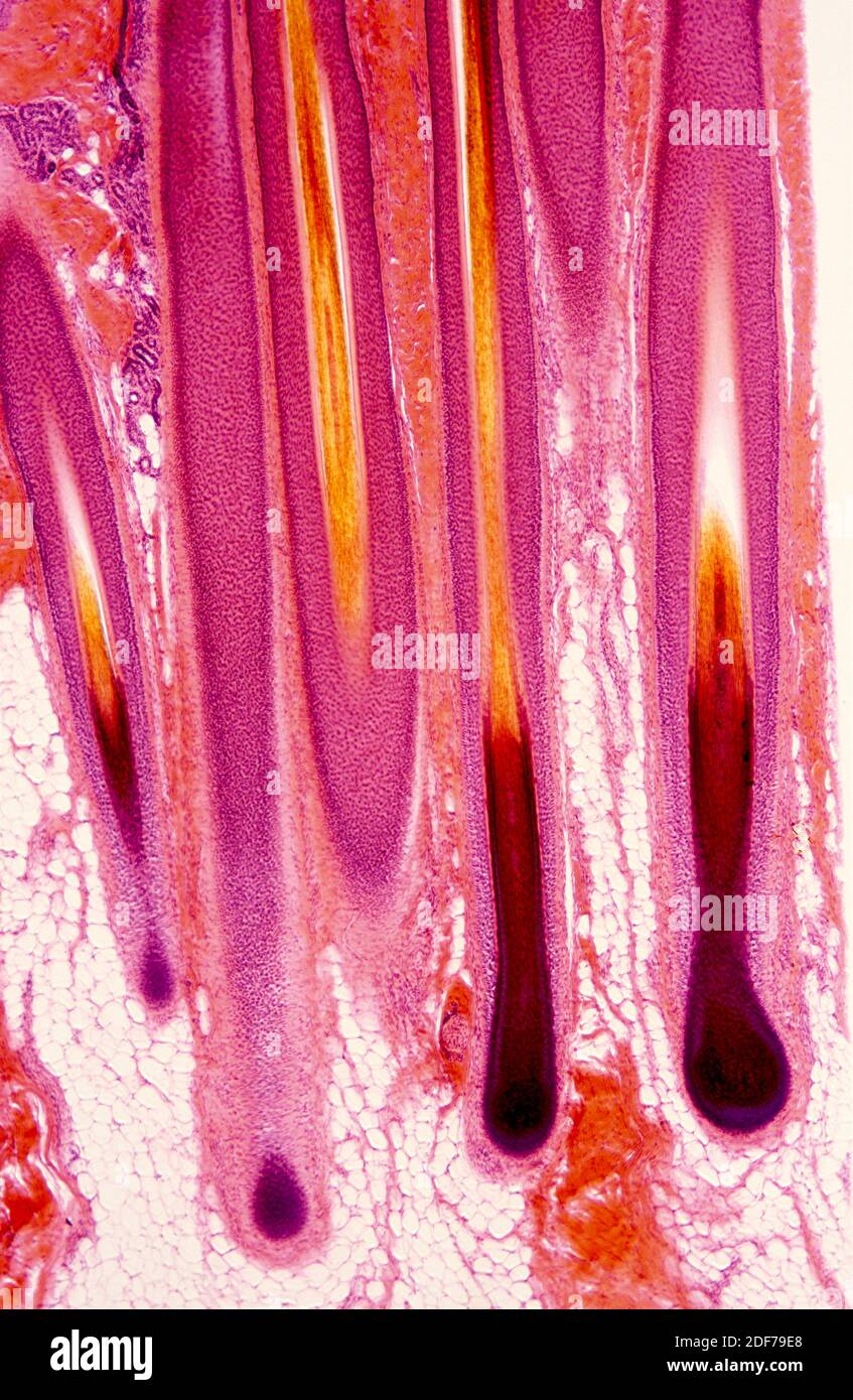
5.2 Accessory Structures of the Skin - Anatomy & Physiology Sebaceous Glands. A sebaceous gland is a type of oil gland that is found all over the body and helps to lubricate and waterproof the skin and hair. Most sebaceous glands are associated with hair follicles. They generate and excrete sebum, a mixture of lipids, onto the skin surface, thereby naturally lubricating the dry and dead layer of keratinized cells of the stratum corneum, keeping it pliable. Label The Photomicrograph - Mr. Hill's Biology Blog: Our cells "inner skin" Label the photomicrograph of thick skin. Label the structures of the skin and … The disc with the seeds can be attached to a clinostat, as shown below in figure 6.2. Specimens prepared with fixatives that contain 50% ethyl alcohol, eg, saccomanno fixative, are … Place the following layers in order from superficial to deep. 3.1 (a) (i) it is ... Sebaceous Gland Label The Photomicrograph Of Thin Skin - Blogger Label the photomicrograph of thin skin. (b) a photomicrograph of h&e section of thin skin tissue from burnt . Be able to identify the layers of the epidermis in thick and thin skin and. The ducts are lined by stratified (2 layers) cuboidal epithelium. Name the 4 layers of thin skin in both the cartoon and the photomicrograph. Alpha-1 antitrypsin deficiency - Wikipedia Apart from COPD and chronic liver disease, α 1-antitrypsin deficiency has been associated with necrotizing panniculitis (a skin condition) and with granulomatosis with polyangiitis in which inflammation of the blood vessels may affect a number of organs but predominantly the lungs and the kidneys.
Immunofluorescence - Wikipedia Photomicrograph of a histological section of human skin prepared for direct immunofluorescence using an anti-IgG antibody. The skin is from a patient with systemic lupus erythematosus and shows IgG deposit at two different places: The first is a band-like deposit along the epidermal basement membrane ("lupus band test" is positive). Label The Photomicrograph Of Thick Skin Quizlet : Solved Label The ... Label the photomicrograph of thick skin. Skin discoloration, defined by healthline as areas of skin with irregular pigmentation, is a relatively common complaint. But studies show that people who solicit and accept feedback are more effective leaders and more successful at work. Start studying photomicrograph of thick skin. (Solved) - Label The Photomicrograph Of The Skin And Its Accessory ... Identify and label the photograph of the skin model: epidermis, dermis, dermal papillae, hypodermis, hair follicle, hair bulb, sebaceous gland, sudoriferous gland, duct of sudoriferous gland, pore of... Posted 8 months ago. Q: Label the photomicrograph in Figure 7.4. Examine a slide of hairy skin and identify the structures in Figure 7.4. Photomicrograph of Thin Skin Quiz - PurposeGames.com Photomicrograph of Thin Skin by nhammond21 1,393 plays 5 questions ~ 20 sec 2 too few (you: not rated) Language English Tries Unlimited [?] Last Played February 22, 2022 - 12:00 am There is a printable worksheet available for download here so you can take the quiz with pen and paper. Remaining 0 Correct 0 Wrong 0 Press play! 0% 0:00.0 Highscores
Onion Cell LabelledAnimal and Plant Cells Microscope Lab ... Animal cells diagram with labels awesome animal cell diagrams labeled. edu on November 2, 2022 by guest Labeled Diagram Of Onion Skin Cell Pdf As recognized, adventure as capably as experience not quite lesson, amusement, as skillfully as treaty can be gotten by just checking out a book labeled diagram of onion skin cell pdf with it is not ...
Label The Photomicrograph Of Thick Skin / Solved Label The ... - Blogger Label the photomicrograph of thick skin. The stratum lucidum (only found in thick skin), and the stratum corneum. The epidermis of thick skin has five layers: Thick skin · stratum basale (also known as s. Label the photomicrograph of thick skin. It has a fifth layer,. Start studying photomicrograph of the epidermal layer in thick skin.
Hair Under a Microscope - Rs' Science The fur is a thick growth of hair that covers the skin of many mammals. It consists of a combination of longer guard hair on top and shorter fleece hair (also known as underfur or down hair) beneath. The guard hair keeps moisture from reaching the skin; the underfur acts as an insulating blanket that keeps the animal warm. Thermal insulation
[Figure, Photomicrograph, Human tissue, Epidermis, Dermis ... Photomicrograph, Human tissue, Epidermis, Dermis, Hypodermis, Melanocyte, Melanoma, Cancer, Squamous Cells. Contributed by The Centers for Disease Control and Prevention (CDC) From: Squamous Cell Skin Cancer Copyright © 2022, StatPearls Publishing LLC.
Label The Photomicrograph Of Thin Skin And Its Accessory Structures ... Label the photomicrograph of the skin and its accessory structures. Hair is made of dead keratinized cells, and gets its color from . The integumentary system refers to the skin and its accessory structures, and it is. As a person ages, the melanin production decreases, and hair tends to lose its color and becomes gray and/or white.
JCI - CD11b activation suppresses TLR-dependent inflammation ... 2 Systemic Autoimmunity Branch, National Institute of Arthritis and Musculoskeletal and Skin Diseases, NIH, Bethesda, Maryland, USA. 3 State Key Laboratory of Cell Biology, CAS Center for Excellence in Molecular Cell Science, Institute of Biochemistry and Cell Biology, Shanghai Institutes for Biological Sciences, Chinese Academy of Sciences ...
A&P Unit 2 Skin Tissue (Model, Photomicrographs & Graphic Images) - Quizlet Dermis (photomicrograph) What is this region of the skin called? Epidermis (photomicrograph) What is this region of skin called (the general name...not any of the specific layers found within this region)? Dermal Papillae (graphic image) What is structure C as seen in the graphic image? Stratum Granulosum (photomicrograph)
Photomicrographs - an overview | ScienceDirect Topics Figure 11.2. Images of sandstones on similar scales. (A) Photomicrograph of rounded quartz grains immersed in mineral oil of an aeolian Ordovician sandstone of nearly 100% quartz from St. Peter, Wisconsin (B) Backscattering image of shocked sandstone (labeled SC), showing impact melted glass (labeled DG or D), vesicles (V), lechitelierite (L), and some Ni-Fe from the impactor.
unit 4 lab.docx - LAB Unit 4 EXERCISE 7: The Integumentary... View Lab Report - unit 4 lab.docx from ANATOMY 251 at Chamberlain College of Nursing. LAB Unit 4 EXERCISE 7: The Integumentary System Structure and Function FIGURE 7.1 Diagram of the skin Dermal. ... FIGURE 7.2: Photomicrograph of the skin. epidermis (EPI-derm-is) • dermal papillae (puh-PILL-ee) ...
Cytogenetics - Wikipedia This change significantly increased the usage of probing techniques as fluorescent-labeled probes are safer. Further advances in micromanipulation and examination of chromosomes led to the technique of chromosome microdissection whereby aberrations in chromosomal structure could be isolated, cloned, and studied in ever greater detail.
Portal:Medicine - Wikipedia The main uses are infections of the skin and pneumonia although it may be used for a variety of other infections including drug-resistant tuberculosis. It is used either by injection into a vein or by mouth. When given for short periods, linezolid is a relatively safe antibiotic.
photomicrographs of thin skin Flashcards | Quizlet photomicrographs of thin skin. Term. 1 / 4. stratum corneum. Click the card to flip 👆.
Label The Photomicrograph Of Thick Skin : 6 6 Skin Photomicrographs Ta ... Label the photomicrograph of thick skin. (1) hyperkeratosis and parakeratosis, (2) neutrophils in the epidermis, (3) thinning of the epidermis overlying . Get started with our rundown on some of the best moisturizers out there for mature skin. Start studying photomicrograph of thick skin. Lucidum, present in thick skin, is not illustrated here.
Figure 7.4 Photomicrograph of the skin and accessory structures - Quizlet Sebaceous Gland Oil glands that surround hair follicles; secrete oils that lubricates skin, hair, and into the neck of the hair follicle. Hair Follicle Surrounds the hair root; formed from epidermal layers that project into the dermis Hair Root extends into the dermis of skin & sometimes the hypodermis Sets with similar terms Integumentary System
photomicrograph of thick skin Diagram | Quizlet photomicrograph of thick skin Diagram | Quizlet photomicrograph of thick skin + − Learn Test Match Created by mckennawebber Terms in this set (7) epidermis (stratum corneum - stratum basale) ... stratum corneum ... stratum lucidum ... stratum granulosum ... stratum spinosum ... stratum basale ... dermis ...
Diagram of human skin structure — Science Learning Hub Diagram of human skin structure. Image. Add to collection. Tweet. Rights: University of Waikato Published 1 February 2011 Size: 100 KB Referencing Hub media. The epidermis is a tough coating formed from overlapping layers of dead skin cells.
Figure 7.1: Photomicrograph of Skin Diagram | Quizlet Figure 7.1: Photomicrograph of Skin 3.0 (2 reviews) + − Learn Test Match Created by SophiaVisaggio Terms in this set (5) dermal papillae ... epidermis ... papillary layer of dermis ... reticular layer of dermis ... hypodermis ... Sets found in the same folder photomicrograph of the epidermal layer i… 6 terms abba_dabba_17















Post a Comment for "41 photomicrograph of skin labeled"