40 art-labeling activity: structural organization of skeletal muscle
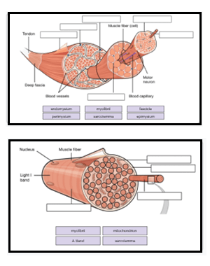
PDF In this chapter, you will learn that - Pearson Each skeletal muscleis a discrete organ, made up of several kinds of tissues. Skeletal muscle fibers predominate, but blood vessels, nerve fibers, and substantial amounts of connective tissue are also present. We can easily examine a skeletal muscle's shape and its attachments in the body without a microscope. Nerve and Blood Supply Art-labeling Activity: Structure of Compact Bone - Quizlet Start studying Art-labeling Activity: Structure of Compact Bone. Learn vocabulary, terms, and more with flashcards, games, and other study tools.
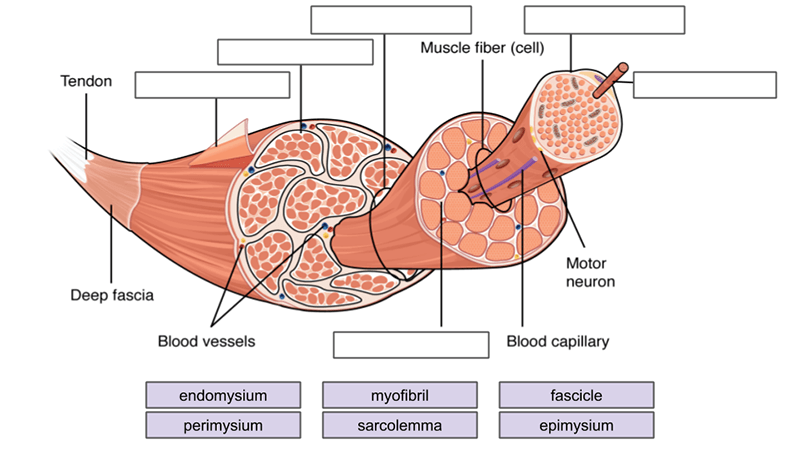
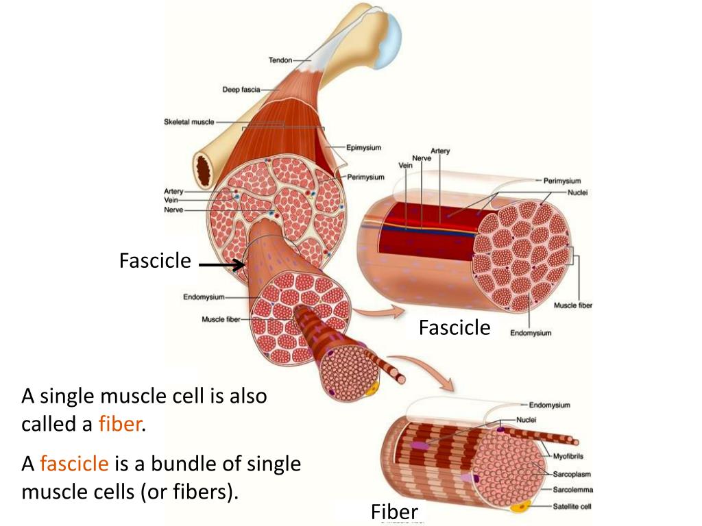
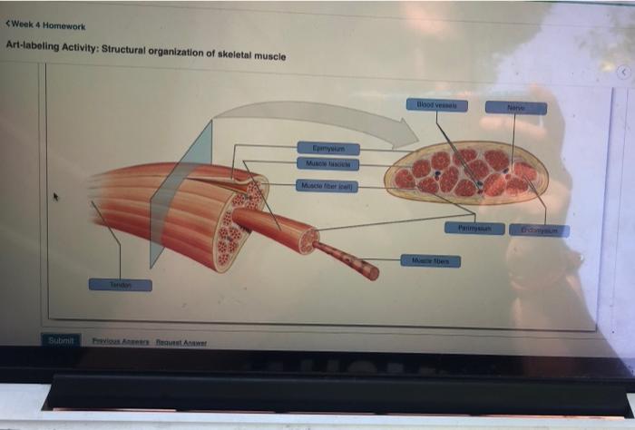
BIOL.docx - Ch9 Hmwk Art-labeling Activity: Structural organization of ... Art-labeling Activity: Structural organization of skeletal muscle Part A Drag the labels to the appropriate location in the figure. Epimysium Muscle fascicle Endomysium Perimysium Nerve Muscle fibers Blood vessels Tendon Muscle fiber (cell) Help Reset Submit My Answers Give Up Correct Provide FeedbackContinue

Art-labeling activity: structural organization of skeletal muscle
Answered: Art-labeling Activity: Structural… | bartleby Answered: Art-labeling Activity: Structural… | bartleby. Homework help starts here! Science Biology Q&A Library Art-labeling Activity: Structural organization of skeletal muscle Reset Epimysium Muscle fascicle Endomysium Perimysium Nerve Muscle fibers Blood vessels Tendon Muscle fiber (cell) clarice-langhans Art Labeling Activity: The Structure Of A Sarcomere : Solved: Art-labeling Activity: Cross Section Of A Skeletal... | Chegg.com - Figure as an art labeling activity. Sarcomere (contractile unit) of a myofibril. Structural organization of skeletal muscle reset epimysium muscle fascicle endomy... Solved Expert Answer 100% (1 rating) Structural organisation of skeletal muscle 1.The part tendon is labelled correctly in the diagram The tendon is a soft tissue by which muscle attaches to bone 2.The part epimysium is labelled correctly The dense connective tissue that su … View the full answer
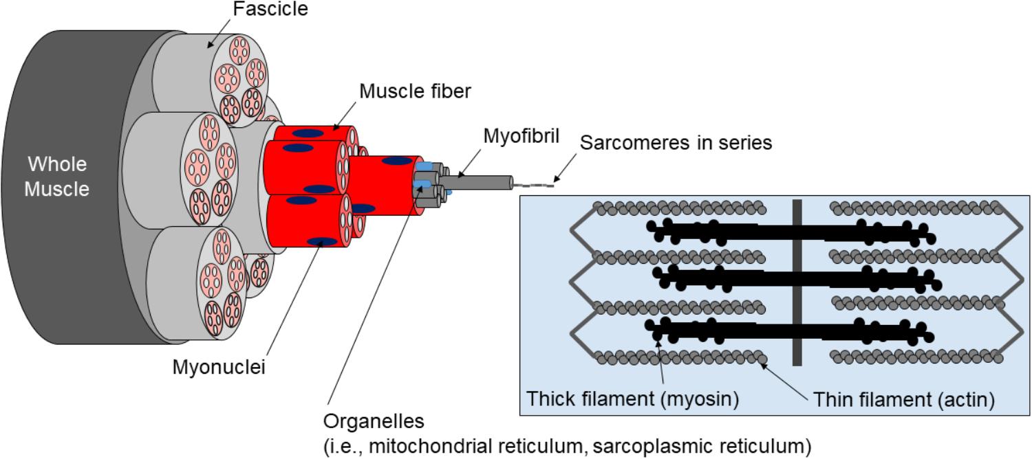
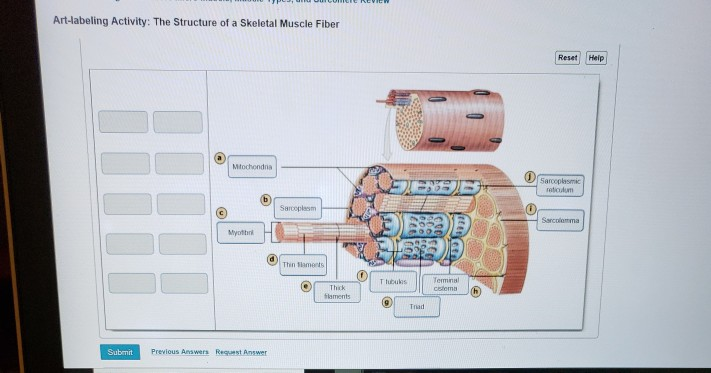
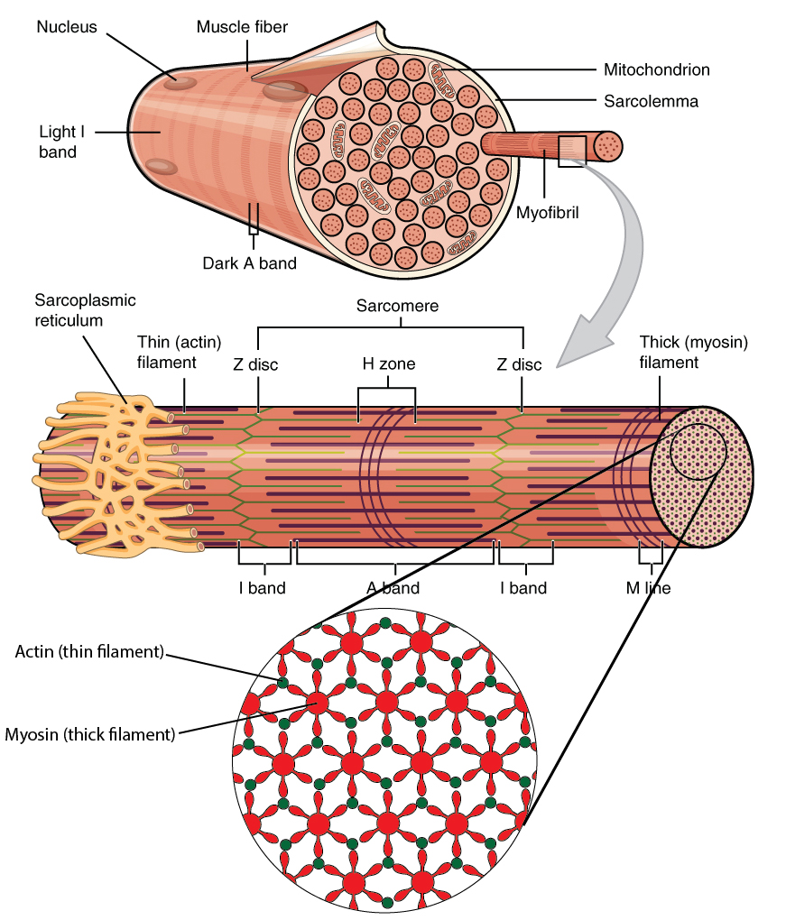
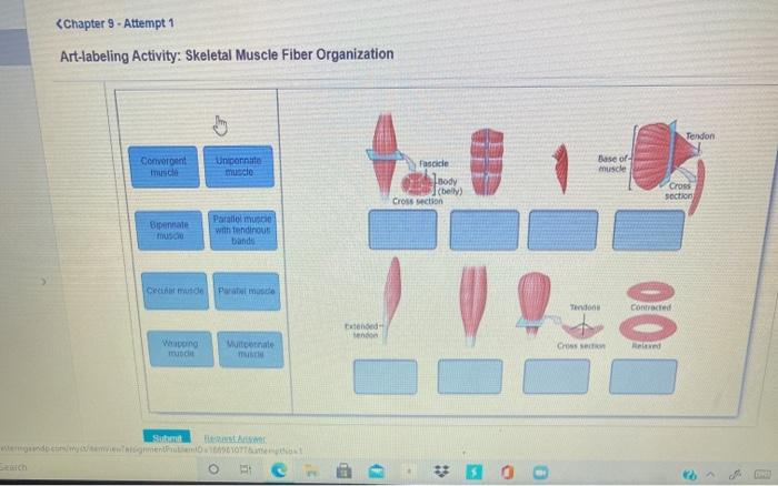
Art-labeling activity: structural organization of skeletal muscle. Week 3 Chapter 9.pdf - 4/23/22, 5:03 PM Week 3 Chapter 9... - Course Hero 4/23/22, 5:03 PM Week 3 Chapter 9 6/10 Label the various arrangements of skeletal muscle fibers. Part A Drag the correct label to the appropriate location to identify the various arrangements of skeletal muscle fibers. ANSWER: Correct Spotlight Figure 9.13: Levers and Pulleys Read through Spotlight Figure 9.13, and then complete the questions and activity below. To label: The muscle types given according to its fascicle organization ... To label: The muscle types given according to its fascicle organization. Introduction: The muscular system consists of skeletal, smooth, and cardiac muscles. This system contributes more to the body weight than other organ system of the human body. It allows the body movement, maintains posture, and helps in blood circulation throughout the body. Art-labeling Activity: The Structure of a Skeletal Muscle Fiber Start studying Art-labeling Activity: The Structure of a Skeletal Muscle Fiber. Learn vocabulary, terms, and more with flashcards, games, and other study tools. Skeletal Muscle Fiber Structure and Function - Open Textbooks for Hong Kong Each skeletal muscle fiber is a skeletal muscle cell. Within each muscle fiber are myofibrils, long cylindrical structures that lie parallel to the muscle fiber. Myofibrils run the entire length of the muscle fiber. They attach to the plasma membrane, called the sarcolemma, at their ends, so that as myofibrils shorten, the entire muscle cell ...
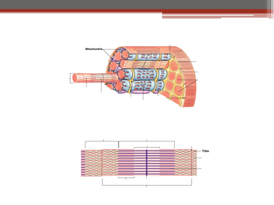
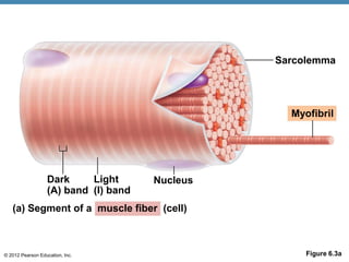
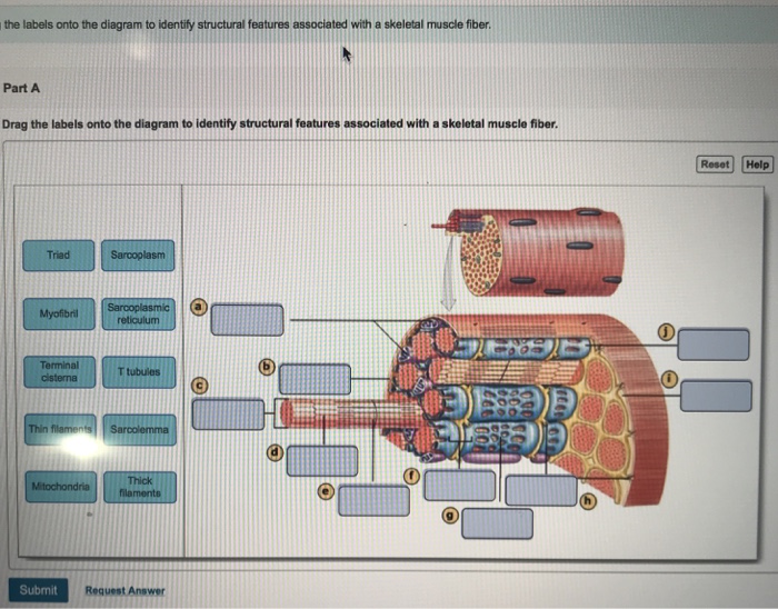
Part a muscle tissue has the ability to contract when Correct Art-labeling Activity: The Organization of Skeletal Muscles (2 of 2) Drag the labels onto the diagram to identify structural features associated with skeletal muscle. Part A Drag the labels onto the diagram to identify structural features associated with skeletal muscle. PDF Screen Shot 2019-03-23 at 10.48.50 AM - Los Angeles Mission College Part of a skeletal muscle fiber (cell) Sarcolemma Microscopic Anatomy and Organization of Skeletal MUSC e 185 I band Z disc Myofibril A band H zone line I band Z disc Sarcolemma Triad: T tubule Terminal cisterns of the SR (2) Tubules of the SR Myofibrils Mitochondria Instructors may assign this figure as an Art Labeling Activity using Mastering A&P 1- CHAPTER 9 MASTERING ASSIGNMENTS Flashcards | Quizlet PICTURE Art-labeling Activity: The structure of a skeletal muscle fiber PICTURE Which thin filament-associated protein binds two calcium ions? troponin Action potential propagation in a skeletal muscle fiber ceases when acetylcholine is removed from the synaptic cleft. A&P. Anatomy and physiology. Pearson labs. - StuDocu Part A In both human and the sheep brain, the cerebellum is the most prominent structure. ANSWER: Correct. Art-labeling Activity: Internal Structures of a Midsagittal Section of the Brain. Part A Drag the labels to the appropriate location in the figure. ANSWER: Correct. Lab Manual Exercise 15 Pre-lab Quiz Question 5
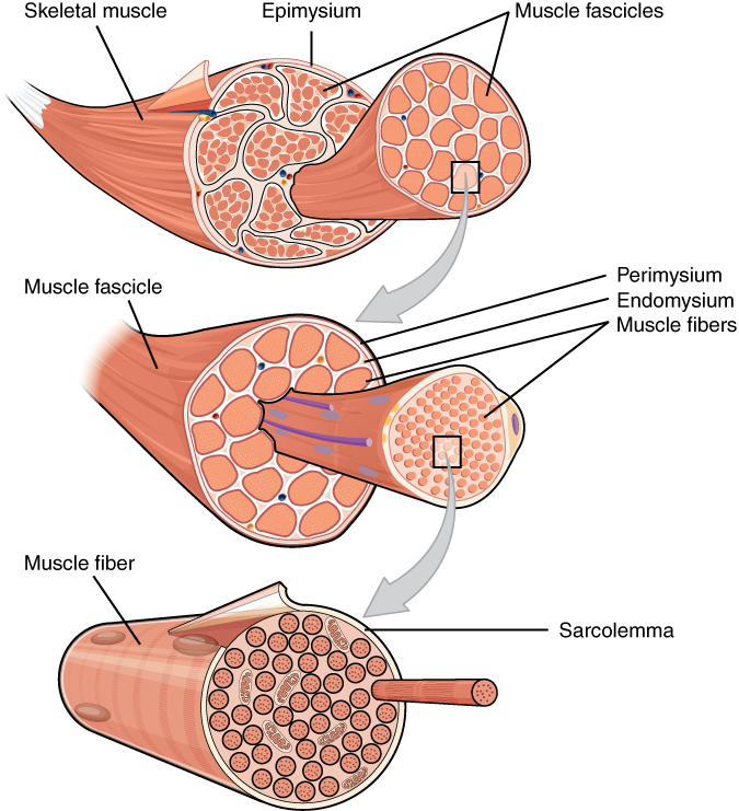
10.2 Skeletal Muscle - Anatomy and Physiology 2e | OpenStax Skeletal muscles are located throughout the body at the openings of internal tracts to control the movement of various substances. These muscles allow functions, such as swallowing, urination, and defecation, to be under voluntary control. art-labeling activity: bones of the axial skeleton - woodrow-grieves Exercise 9 Review Sheet Art-labeling Activity 1 1 of 3 25 of 50 Part A Drag the labels onto the diagram to identify the bones of the skull. Point where the hip bone that receives the head of the thigh bone. Lab re ee exercise lab 10 the appendicular skeleton bones of the pectoral girdle and upper limb 1. An answer key is not. Art-labeling Activity: Long Section of a Skeletal Muscle Start studying Art-labeling Activity: Long Section of a Skeletal Muscle. Learn vocabulary, terms, and more with flashcards, games, and other study tools. Access Free Chapter 9 Muscle Tissue Answer Key Chapter Games and Activities Art-Labeling Activities Connective tissue sheaths of skeletal muscle: epimysium, perimysium, and endomysium (Figure 9.1) Microscopic anatomy of a skeletal muscle fiber (Figure 9.2a-b) Microscopic anatomy of a skeletal muscle fiber (Figure 9.2c-d) Study Guide p. 189-197 KEY - Chapter 9 Muscles and Muscle ...
Human anatomy and physiology, Basic anatomy and physiology ...
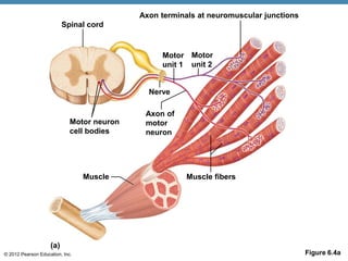
BSC2085L Chapter 013 Activity 1 Skeletal Muscle Organization 005 Part A ... BSC2085L Chapter 013 Activity 1 Skeletal Muscle Organization-005 Part A The area of a sarcomere where the thin actin filaments connect to one another is called the _____. ANSWER: Correct The Z line or Z disc consists of proteins called actinin that anchor the actin filaments together. A message from your instructor... Activity 2: The Neuromuscular Junction Art-labeling Activity: Skeletal ...
Muscular Levels of Organization | Anatomy and Physiology I ...
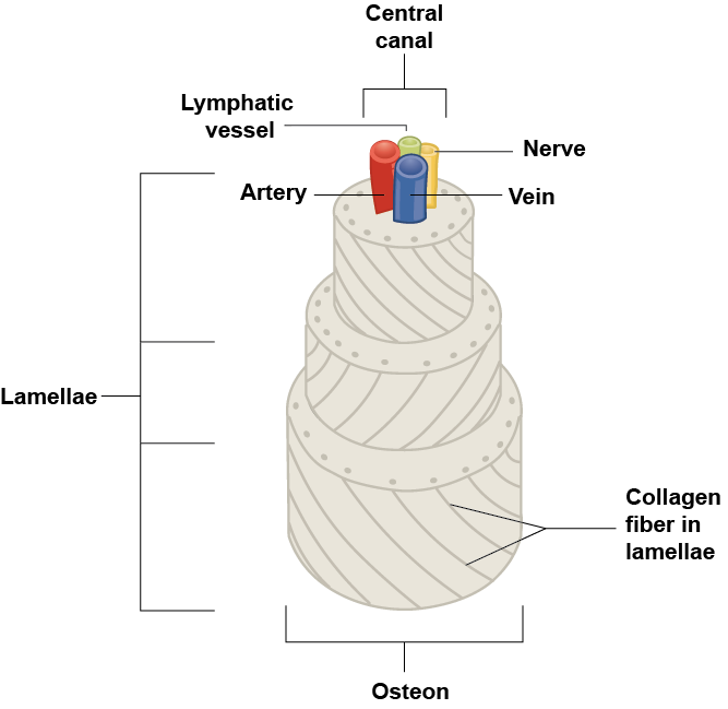
Anatomy 260 Ch3 copy.pdf - Week 1 Chapter 3_ Due: 10:59pm... - Course Hero Correct Art-labeling Activity: Anatomy and Histological Organization of Bone Label the structural features of compact bone. Part A Drag the labels to the appropriate location in the figure. ANSWER: Help Reset Apical portion of cell breaking down Golgi apparatus Secretory vesicles fusing with the plasmalemma Stem cell dividing to replace lost ...

components and divisions of the pelvis.jpg - ring A&P ...
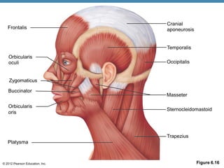
Muscle Review: Labeling Activities - pearsoncmg.com Test your visual understanding of body structures with these interactive labeling activities. Muscles of facial expression Muscles of the vertebral column - posterior view of superficial muscle layer Muscles of the vertebral column - anterior surfaces of the superior vertebrae Muscles that position the pectoral girdle - posterior view

Anatomy: Chapter 9 Skeletal Muscular System LABELING ...
Art- labeling Activity Flashcards | Quizlet Study with Quizlet and memorize flashcards containing terms like , , and more.
.jpg)
Chapter 38 - Skeletal Muscle - BIO 140 - Human Biology I ...
Pre-Lab Microscopic Anatomy ans Organization of Muscles - StuDocu Microscopic Anatomy and Organization of Skeletal Muscle Learning Outcomes Define muscle fiber, myofibril , and myofilament, and describe the structural relationships among th em. Describe thick (myosin) and thin (actin) filaments and their relationship to the sarcomere. Discuss the struct ure and location of T tubules and terminal cisterns t- Define endomys ium, p erimysium, and epimysium, and ...
Frontiers | A Critical Evaluation of the Biological Construct ...
Solved Expert Answer 100% (1 rating) Structural organisation of skeletal muscle 1.The part tendon is labelled correctly in the diagram The tendon is a soft tissue by which muscle attaches to bone 2.The part epimysium is labelled correctly The dense connective tissue that su … View the full answer

BIO 200 Chapter 9 - Muscle Tissue Physiology Flashcards | Quizlet
clarice-langhans Art Labeling Activity: The Structure Of A Sarcomere : Solved: Art-labeling Activity: Cross Section Of A Skeletal... | Chegg.com - Figure as an art labeling activity. Sarcomere (contractile unit) of a myofibril. Structural organization of skeletal muscle reset epimysium muscle fascicle endomy...

Secretome Analysis of Lipid-Induced Insulin Resistance in ...
Answered: Art-labeling Activity: Structural… | bartleby Answered: Art-labeling Activity: Structural… | bartleby. Homework help starts here! Science Biology Q&A Library Art-labeling Activity: Structural organization of skeletal muscle Reset Epimysium Muscle fascicle Endomysium Perimysium Nerve Muscle fibers Blood vessels Tendon Muscle fiber (cell)
Muscles Labeling

Muscular Levels of Organization | Anatomy and Physiology I ...
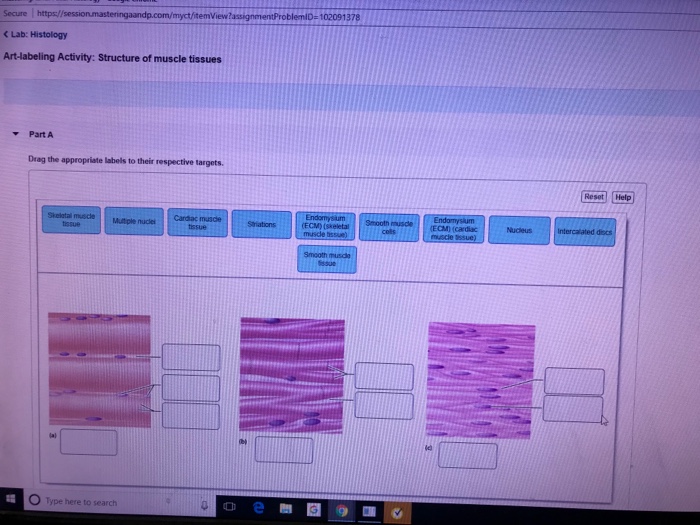
Solved Secure https:/ C Lab: Histology Art-labeling | Chegg.com

Week 6: Muscle Physiology Flashcards | Quizlet
A & P Ch 6 Musclular System Student PPT
Microscopic Anatomy and Organization of Skeletal Muscle

Chapter 38 - Skeletal Muscle - BIO 140 - Human Biology I ...
Muscles and Muscle Tissue

9 The Muscular System: Skeletal Muscle Tissue and ...
Solved Art-labeling Activity: The Structure of a Skeletal ...
SKELETAL MUSCLE ORGANIZATION
Muscles Labeling
6.3 Bone Structure – Anatomy & Physiology

Art-labeling Activity: Long Section of a Skeletal Muscle ...
Human Anatomy & Physiology Ninth Edition
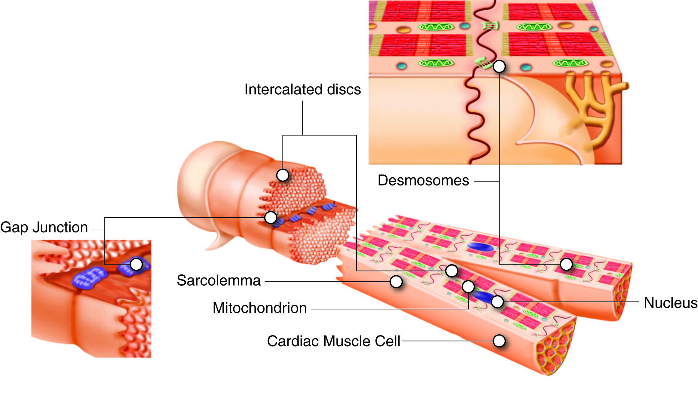
The Cardiovascular System —
Skeletal Muscle - Etsy
PPT - Muscle Tissue PowerPoint Presentation, free download ...
Chapter 10: Muscle Tissue. Muscle Tissue A primary tissue ...
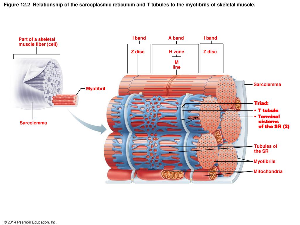
Figure 12.1 Microscopic anatomy of skeletal muscle. - ppt ...

Ex 12 Microscopic Anatomy & Organization of Skeletal Muscle ...

Muscular Levels of Organization | Anatomy and Physiology I ...
OVERVIEW OF MUSCLE TISSUE
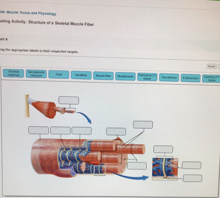
Solved Art-labeling activity: structure of skeletal muscle ...
A & P Ch 6 Musclular System Student PPT
A & P Ch 6 Musclular System Student PPT

1.2 Structural Organization of the Human Body – Anatomy ...
Solved the labels onto the diagram to identify structural ...

MHC is critical for muscle striation formation. (A) Schematic ...
Solved
9.17: Skeletal Muscle - Medicine LibreTexts
Solved
Post a Comment for "40 art-labeling activity: structural organization of skeletal muscle"