40 compound microscope diagram
Compound Microscope Diagram Diagram | Quizlet Connects the nosepiece to the rest of the microscope. Stage. Holds the slide being viewed. Coarse Adjustment Knob. Used to bring a specimen into view by making large adjustments to the stage. Focus. Being able to see a specimen clearly. Fine Adjustment Knob. Helps to focus your view of a specimen by making small adjustments the the stage. Diagram of a Compound Microscope - Biology Discussion A bright-field or compound microscope is primarily used to enlarge or magnify the image of the object that is being viewed, which can not otherwise be seen by the naked eye. Magnification may be defined as the degree of enlargement of the image of an object provided by the microscope.
Compound Microscope- Definition, Labeled Diagram, Principle, … Apr 03, 2022 · Magnification of compound microscope. In order to ascertain the total magnification when viewing an image with a compound light microscope, take the power of the objective lens which is at 4x, 10x or 40x and multiply it by …

Compound microscope diagram
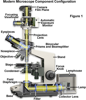
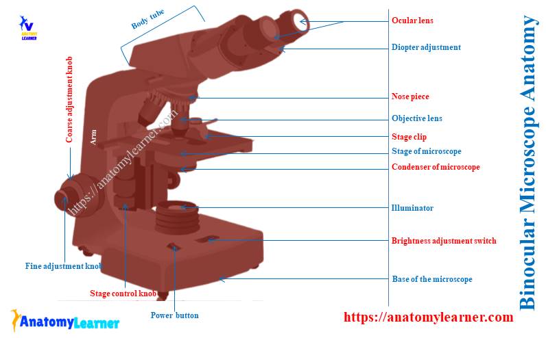
microscope | Types, Parts, History, Diagram, & Facts Aug 22, 2022 · The most familiar type of microscope is the optical, or light, microscope, in which glass lenses are used to form the image. Optical microscopes can be simple, consisting of a single lens, or compound, consisting of several optical components in line. The hand magnifying glass can magnify about 3 to 20×. Single-lensed simple microscopes can ... Compound Microscope Parts - Labeled Diagram and their Functions There are three major structural parts of a compound microscope. The head includes the upper part of the microscope, which houses the most critical optical components, and the eyepiece tube of the microscope. The base acts as the foundation of microscopes and houses the illuminator. The arm connects between the base and the head parts. Working Principle and Parts of a Compound Microscope (with Diagrams) ADVERTISEMENTS: Read this article to learn about the working principle and parts of a compound microscope with diagrams! Working Principle: The most commonly used microscope for general purposes is the standard compound microscope. It magnifies the size of the object by a complex system of lens arrangement. ADVERTISEMENTS: It has a series of two lenses; […]
Compound microscope diagram. Compound Microscope Parts, Function, & Diagram - Study.com The definition of a compound microscope is "an upright microscope that utilizes two different lenses to magnify the size of the objects being viewed." The name itself describes what it is. The tern... Microscopy: History, Types of Microscope, Diagram - Embibe Compound Microscope. Definition: Compound microscopes are dual-lens microscopes. The two lenses are called objective lens and eyepiece. It is commonly referred to as a student microscope. Lens used: A compound microscope uses two convex lenses, one in the eyepiece and another in the objective. The eyepiece lenses vary with their focal lengths and hence the magnifications also differ. Parts of a Compound Microscope (And their Functions) - Scope Detective A compound microscope is the most common microscope you can get and the type you'll typically see in a lab or hobbyist's study. These microscopes tend to have total magnification between 40x - 2000x to allow you to see specimens like bacteria and cells. Microscope, Microscope Parts, Labeled Diagram, and Functions Jan 19, 2022 · Revolving Nosepiece or Turret: Turret is the part of the microscope that holds two or multiple objective lenses and helps to rotate objective lenses and also helps to easily change power. Objective Lenses: Three are 3 or 4 objective lenses on a microscope. The objective lenses almost always consist of 4x, 10x, 40x and 100x powers. The most common eyepiece …
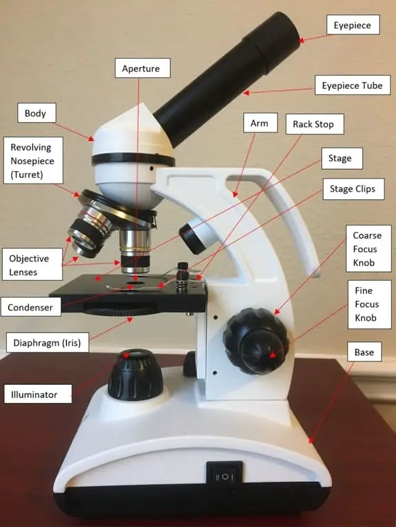
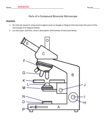
Microscope Parts and Functions Microscope Parts and Functions With Labeled Diagram and Functions How does a Compound Microscope Work?. Before exploring microscope parts and functions, you should probably understand that the compound light microscope is more complicated than just a microscope with more than one lens.. First, the purpose of a microscope is to magnify a … Microscope: Types of Microscope, Parts, Uses, Diagram - Embibe A compound microscope is defined as a microscope with a high resolution. It uses two sets of lenses, providing a \ (2\)-dimensional image of the sample. The term compound refers to the usage of more than one lens in the microscope. Also, the compound microscope is one of the types of optical microscopes. Microscope Parts and Functions First, the purpose of a microscope is to magnify a small object or to magnify the fine details of a larger object in order to examine minute specimens that cannot be seen by the naked eye. Here are the important compound microscope parts... Eyepiece: The lens the viewer looks through to see the specimen. Compound Microscope: Definition, Diagram, Parts, Uses, Working ... - BYJUS The parts of a compound microscope can be classified into two: Non-optical parts Optical parts Non-optical parts Base The base is also known as the foot which is either U or horseshoe-shaped. It is a metallic structure that supports the entire microscope. Pillar The connection between the base and the arm are possible through the pillar. Arm
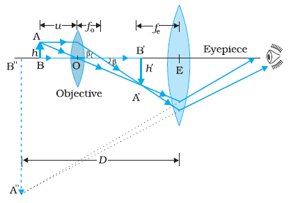
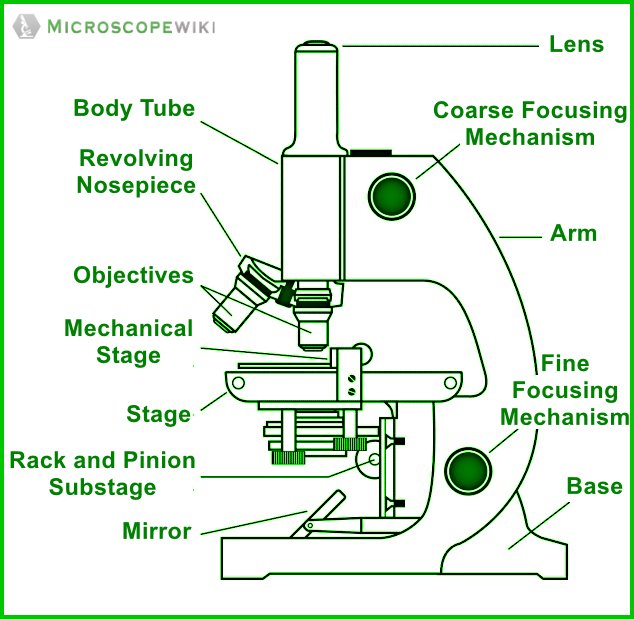
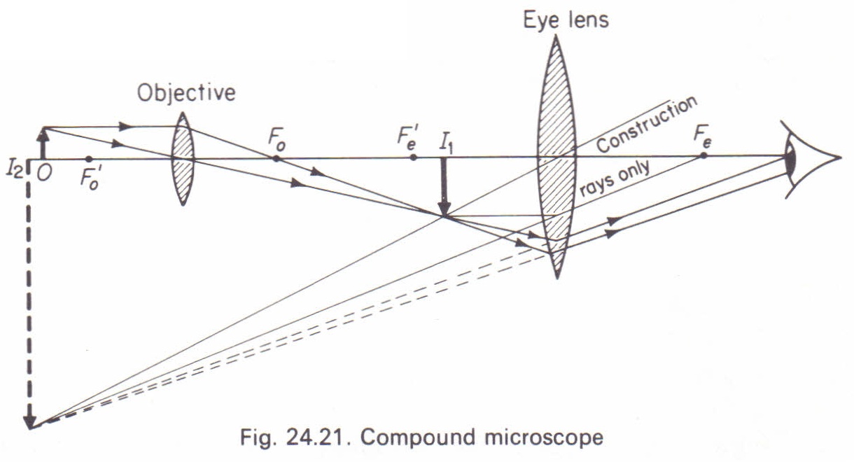
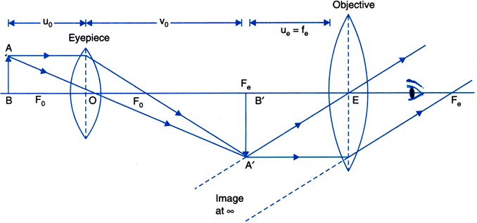
A Study of the Microscope and its Functions With a Labeled Diagram ... These labeled microscope diagrams and the functions of its various parts, attempt to simplify the microscope for you. However, as the saying goes, 'practice makes perfect', here is a blank compound microscope diagram and blank electron microscope diagram to label. Download the diagrams and practice labeling the different parts of these ... Parts of a Compound Microscope and Their Functions - NotesHippo Compound microscope mechanical parts (Microscope Diagram: 2) include base or foot, pillar, arm, inclination joint, stage, clips, diaphragm, body tube, nose piece, coarse adjustment knob and fine adjustment knob. Base: It's the horseshoe-shaped base structure of microscope. All of the other components of the compound microscope are supported ... The compound microscope - how to draw ray diagrams - YouTube An animated presentation showing you how to draw ray diagrams (using simple lens rules) for a compound microscope. This shows how to determine the position a... Compound Microscope – Diagram (Parts labelled), Principle and … Feb 03, 2022 · The three structural components include: 1. Head – This is the upper part of the microscope that houses the optical parts 2. Arm – This part connects the head with the base and provides stability to the microscope. Arm is used to carry the microscope around 3. Base – Base is on which the microscope rests and the base houses the illuminator that lights up the …
Compound Microscope Parts, Functions, and Labeled Diagram Compound Microscope Parts, Functions, and Labeled Diagram Parts of a Compound Microscope Each part of the compound microscope serves its own unique function, with each being important to the function of the scope as a whole.
Microscopy- History, Classification, Terms, Diagram - The Biology … Jul 29, 2022 · In 1670, Robert Hooke, an English Chemist, Mathematician, Physicist, and Inventor, improvise the microscope of that time and developed the compound microscope. He first developed the 3 lenses microscope. In 1675, Anton Van Leeuwenhoek ground a glass ball into a convex lens and used it to make a single-lens microscope with 270X magnification.
16 Parts of a Compound Microscope: Diagrams and Video Once you have an understanding of the parts of the microscope it will be much easier to navigate around and begin observing your specimen, which is the fun part! The 16 core parts of a compound microscope are: Head (Body) Arm Base Eyepiece Eyepiece tube Objective lenses Revolving Nosepiece (Turret) Rack stop Coarse adjustment knobs
What is a Compound Microscope? - New York Microscope Company A compound microscope is an instrument that is used to view magnified images of small specimens on a glass slide. It can achieve higher levels of magnification than stereo or other low power microscopes and reduce chromatic aberration. It achieves this through the use of two or more lenses in the objective and the eyepiece.
Bright-field microscope (Compound light microscope) - Diagram (Parts ... Bright-field microscope (Compound light microscope) - Diagram (Parts), Principle, Uses Bright-field Microscope By Editorial Team Last updated on February 9, 2022 A bright-field microscope, also known as a compound light microscope is among the simplest of optical microscopes.
Labelled Diagram of Compound Microscope The below mentioned article provides a labelled diagram of compound microscope. Part # 1. The Stand: The stand is made up of a heavy foot which carries a curved inclinable limb or arm bearing the body tube. The foot is generally horse shoe-shaped structure (Fig. 2) which rests on table top or any other surface on which the microscope in kept.
parts of a microscope diagram Microscope parts science compound knob adjustment coarse labeled diagram biology microscopes light label name tools lab functions does structure 6th. Plant phloem vascular cells anatomy parenchyma primary cambium sclereids fibers tissue sieve elements conducting differentiated procambium initiated secondary called grkraj.
Compound Microscope - Types, Parts, Diagram, Functions and Uses A compound microscope has two convex lenses; an objective lens and eye piece. The objective lens is placed towards the object and the eyepiece is the lens towards our eye. Both eyepiece and objective lenses have a short focal length and fitted at the free ends of two sliding tubes. (4, 5, and 6) Compound microscope parts and magnification
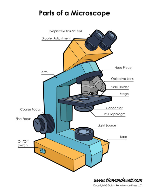
Parts of a microscope with functions and labeled diagram Apr 19, 2022 · Figure: Diagram of parts of a microscope. There are three structural parts of the microscope i.e. head, base, and arm. Head – This is also known as the body. It carries the optical parts in the upper part of the microscope. Base – It acts as microscopes support. It also carries microscopic illuminators.
Compound Light Microscope Diagram Worksheet - Google Groups Modern compound light microscopes under optimal conditions can we an average from 1000X to 2000X times the specimens original diameter Diagram. Label the parts of the microscope using the word...
Label a Compound Microscope Diagram | Quizlet Start studying Label a Compound Microscope. Learn vocabulary, terms, and more with flashcards, games, and other study tools.
Compound Microscope- Definition, Labeled Diagram, Principle, Parts, Uses The naked eye can now view the specimen at magnification 400 times greater and so microscopic details are revealed. Alternatively, the magnification of the compound microscope is given by: m = D/ fo * L/fe where, D = Least distance of distinct vision (25 cm) L = Length of the microscope tube fo = Focal length of the objective lens
Compound Microscope: Parts of Compound Microscope - BYJUS The parts of the compound microscope can be categorized into: Mechanical parts; Optical parts (A) Mechanical Parts of a Compound Microscope. 1. Foot or base. It is a U-shaped structure and supports the entire weight of the compound microscope. 2. Pillar. It is a vertical projection. This stands by resting on the base and supports the stage. 3. Arm
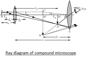
Draw a ray diagram of compound microscope when the class 12 ... - Vedantu Compound Microscope Answer Draw a ray diagram of compound microscope, when the final image is formed at the minimum distance of distinct vision. Answer Verified 202.8k + views Hint: A compound microscope is an optical instrument used for observing highly magnified images of tiny objects.
Compound Microscope - Diagram (Parts labelled), Principle and Uses See: Labeled Diagram showing differences between compound and simple microscope parts Structural Components The three structural components include 1. Head This is the upper part of the microscope that houses the optical parts 2. Arm This part connects the head with the base and provides stability to the microscope.
Parts of a microscope with functions and labeled diagram - Microbe Notes Figure: Diagram of parts of a microscope There are three structural parts of the microscope i.e. head, base, and arm. Head - This is also known as the body. It carries the optical parts in the upper part of the microscope. Base - It acts as microscopes support. It also carries microscopic illuminators.
Parts of Stereo Microscope (Dissecting microscope) – labeled diagram ... If you would like to learn optical components of a compound microscope, please visit Compound Microscope Parts – Labeled Diagram and their Functions, and this article. How to use a stereo (dissecting) microscope. Follow these steps to put your stereo microscopes in work: 1. Set your microscope on a tabletop or other flat sturdy surface where ...
Compound Microscope: Parts of Compound Microscope - BYJUS Diagram Parts of the Compound Microscope Parts of Compound Microscope The parts of the compound microscope can be categorized into: Mechanical parts Optical parts (A) Mechanical Parts of a Compound Microscope 1. Foot or base It is a U-shaped structure and supports the entire weight of the compound microscope. 2. Pillar It is a vertical projection.
Electron Microscope Principle, Uses, Types and Images (Labeled Diagram … Feb 02, 2022 · Ans: A light microscope has a low resolving power (0.25µm to 0.3µm) while the electron microscope has a resolution power about 250 times higher than the light microscope at about 0.001µm. Similarly, a light microscope has a magnification of 500X to 1500x while the electron microscope has a much higher magnification of 100,000X to 300,000X.
Compound Microscope: Know Definition,working, diagram, properties Compound Microscope Diagram. The compound microscope is used to study the structural intricacies of cells, tissues, or organ parts. A compound microscope's components are divided into two categories: Non-optical components. Base: The base, often known as the foot, is either U-shaped or horseshoe-shaped. It is a metallic framework that holds ...
Microscope Parts, Function, & Labeled Diagram - slidingmotion Condenser. The condenser is to focus the light, which passes from the microscopic illuminator to the specimen. This condenser is located just below the diaphragm. This diaphragm is one of the important parts of the compound microscope which will help to get an accurate and sharp image. The condenser has a magnification power of 400X and above.
Working Principle and Parts of a Compound Microscope (with Diagrams) ADVERTISEMENTS: Read this article to learn about the working principle and parts of a compound microscope with diagrams! Working Principle: The most commonly used microscope for general purposes is the standard compound microscope. It magnifies the size of the object by a complex system of lens arrangement. ADVERTISEMENTS: It has a series of two lenses; […]
Compound Microscope Parts - Labeled Diagram and their Functions There are three major structural parts of a compound microscope. The head includes the upper part of the microscope, which houses the most critical optical components, and the eyepiece tube of the microscope. The base acts as the foundation of microscopes and houses the illuminator. The arm connects between the base and the head parts.
microscope | Types, Parts, History, Diagram, & Facts Aug 22, 2022 · The most familiar type of microscope is the optical, or light, microscope, in which glass lenses are used to form the image. Optical microscopes can be simple, consisting of a single lens, or compound, consisting of several optical components in line. The hand magnifying glass can magnify about 3 to 20×. Single-lensed simple microscopes can ...
















Post a Comment for "40 compound microscope diagram"