45 nucleus electron micrograph labelled
Nucleus: Definition, Structure, Functions - Biology Learner The electron micrograph and immunocytological techniques show that three distinct regions are observed in the nucleolus. These are the Fibrillar center, Dense fibrillar component, and Cortical granular components. Fibrillar Center: This pale-staining part represents the innermost region of the nucleolus, which is made up of ribosomal DNA. Electron Micrograph of a Neutrophil - Netter Images Pricing. Price for. Choose Usage Printed publication (book, brochure, journal, etc.) Trial Exhibits and Materials Slide Presentation (Non-web or authenticated login if Web) Electronic Formats Posters Tee Shirts, Novelties Student Lo-res Presentation/Poster, Thesis, Dissertation. Product Description:
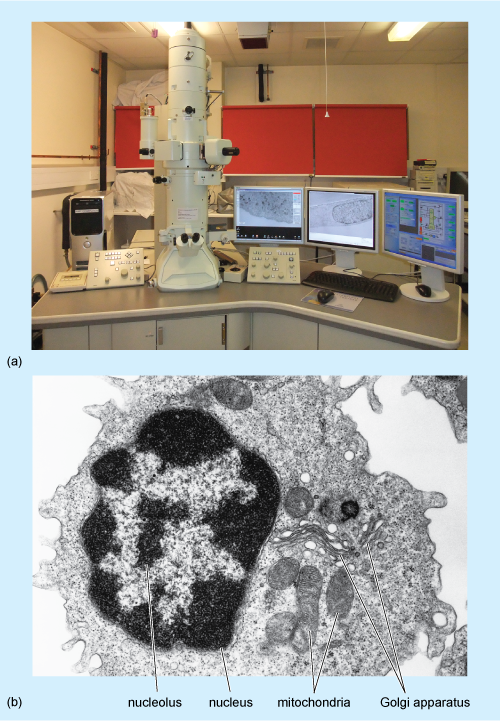
Cell Lab - Yale University The cell's content is divided into two main compartments: the nucleus and the cytoplasm that surrounds the nucleus. Cytoplasm is further divided into organelles, cytosol and inclusions. ... and the electron microscope (magnification up to 500000x). The limit of resolution of the light microscope is 0.2 µm, while the practical limit of ...

Nucleus electron micrograph labelled
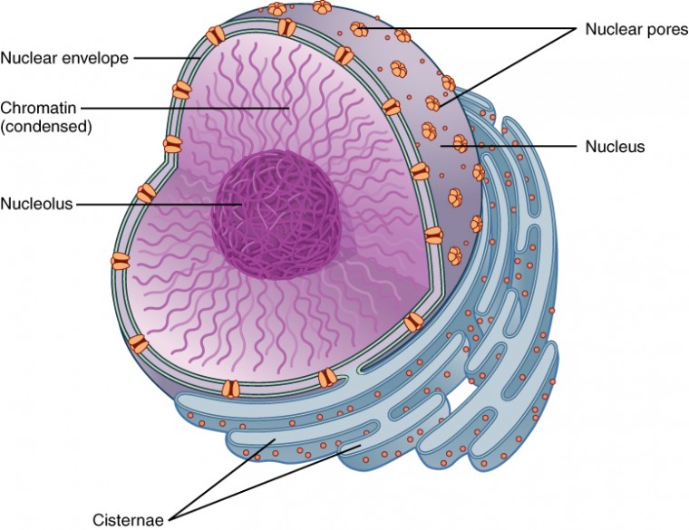
Cell Nucleus - function, structure, and under a microscope Summary. The nucleus is a double-layer membrane organelle. It consists of the nuclear envelope, DNA (chromatin), nucleolus, nucleoplasm, and the nuclear matrix. The main function of the nucleus is to control cell activities and carry genetic information to pass to the next generation. A eukaryotic cell typically has only one nucleus. plant cell label electron micrograph Diagram | Quizlet Start studying plant cell label electron micrograph. Learn vocabulary, terms, and more with flashcards, games, and other study tools. animal cell electron micrograph labelling Diagram | Quizlet Golgi apparatus. ... mitochondria. ... plasma membrane. ... Upgrade to remove ads. Only $2.99/month.
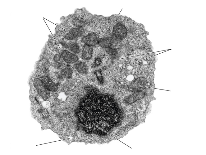
Nucleus electron micrograph labelled. Virtual EM Micrograph List | histology 021. Plasma Cell: This electron micrograph shows a typical secretory cell, a plasma cell, which secretes immunoglobulin protein. Many of the major types of cellular organelles are visible in this image. In the nucleus, areas of euchromatin and heterochromatin can easily be identified. Virtual Slide. Label This Transmission Electron Micrograph : TEM of chloroplast from ... Label the transmission electron micrograph of the nucleus. Provide the labels for the electron micrograph in figure 12.8. Labeling for electron microscopy using antibody conjugated to. Transmission electron microscopy (tem) is a microscopy technique in which a beam of electrons is transmitted through a specimen to form an image. New Tools for Imaging Neutrophils: Work Function Mapping and Element ... Photoemission electron microscopy and imaging X-ray photoelectron spectroscopy are today frequently used to obtain chemical and electronic states, chemical shifts, work function profiles within the fields of surface- and material sciences. Lately, because of recent technological advances, these tools have also been valuable within life sciences. The Cell: The Histology Guide - University of Leeds An electron micrograph of a nucleus Types of Nucleus Cells are normally diploid - this means that they have a pair - two sets of homologous chromosomes, and hence two copies of each gene or genetic locus. However, cells can be haploid, polyploid or aneuploid. Haploid: only has one set of chromosomes - i.e. in a sperm or oocyte.
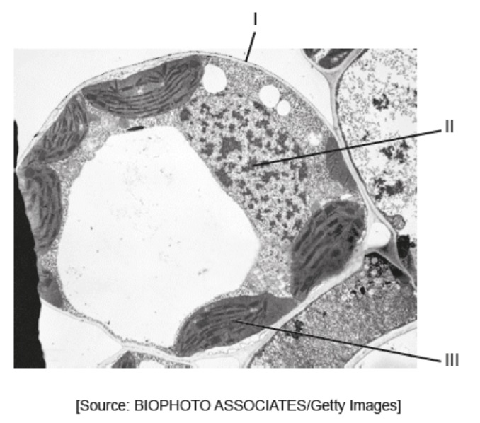
Nucleus - Electron Micrograph Slide 5 of 36 PDF Identifying Organelles from an Electron Micrograph The electron micrograph displayed below illustrates many of the plant cell characteristics discussed The cell wall, large central vacuole and chloroplasts are clearly visible Also visible is the clearly defined nucleus containing chromatin Neuron under Microscope with Labeled Diagram - AnatomyLearner The postsynaptic membrane is an electron density that similar to the presynaptic density. This density reflects the high protein content of the synaptic membrane. Neuron under microscope labelled diagram. Throughout this article, you got the different neurons labelled diagrams. PDF Electron Micrographs (EMs) for laboratories in A215, Basic Human ... - IU Identify and be able to recognize: Label centrioleC The rod-like form of the centriole is seen in longitudinal (lengthwise) section near the right edge of Plate 12a. Note the l-micron scale to the lower left of the large, dark nucleus (N).
The Cell Nucleus Organization | Celebrate Cytochemistry | Gwen V ... Nucleus; The Cell Nucleus Organization ... one can spread these chromosomes on a light microscope slide and label them with dyes that are preferentially taken up by certain regions (for example, modified Giemsa stains). ... up on a plastic-coated grid and examined with the electron microscope. The first level of organization you see is a tangle ... [Immune electron microscope determination of the localization of ... The number of particles observed over diffuse chromatin equals to 50-80% against the label in fibroblast cytoplasm. In contrast, the label used to be absent over the E. coli nucleoid. The presence of TRS in the fibroblast nucleus may evidence in favour of a possible regulatory role of TRS in eukaryots. Label the transmission electron micrograph of the nucleus. Label the transmission electron micrograph of the cell. 0 Nucleus rences Mitochondrion Heterochromatin Peroxisome Vesicle ULAR bumit Click and drag each label into the correct category to indicate whether it pertains to the cytoplasm or the plasma... The Cell Nucleus: A Brief Overview - Microscope Clarity The discovery of electron microscopy was critical in defining the double layered nuclear membrane, nuclear lamina, chromatin packing and nuclear pore complexes. Remarkably, many features of the nucleus including organisation and regulation of genetic material were only elucidated in the 50 years, with new discoveries continuing to be made every ...
Nucleus Electron micrograph Diagram | Quizlet
Ultrastructure and nuclear architecture of telomeric chromatin revealed ... Superimposition of eGFP fluorescence signals and the corresponding EM micrographs from eGFP-APEX2-labelled TRF2 in MEFs, TRF1 in MEFs and TRF1 in U2OS cells demonstrate that telomeres labelled by the APEX2 probes are more electron dense than other chromatin regions in the nucleus (Figure (Figure1C 1C - G).
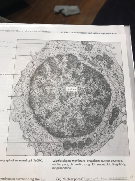
Solved label the ectron micrograph of an animal cell. | Chegg.com
Electron Microscopy - University of Utah Plasma cell. Normal plasma cell with prominent cytoplasmic smooth endoplasmic reticulum. Macrophage. Normal macrophage with oblong nucleus, nucleolus, and cytoplasm with a variety of inclusions. Platelets. Normal platelets. Mitochondria. Happy mitochondria within a cell. Skeletal muscle.
A tour of the cell: View as single page
Electron Micrograph of a Lymphocyte - Netter Images Pricing. Price for. Choose Usage Printed publication (book, brochure, journal, etc.) Trial Exhibits and Materials Slide Presentation (Non-web or authenticated login if Web) Electronic Formats Posters Tee Shirts, Novelties Student Lo-res Presentation/Poster, Thesis, Dissertation. Product Description:

An electron micrograph of a HeLa cell demonstrates that the ...
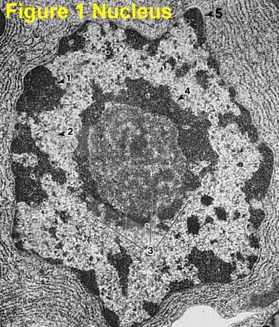
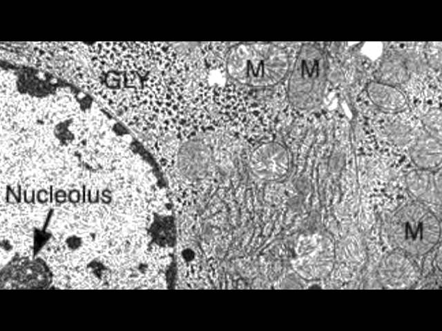
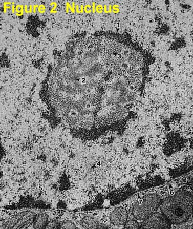
Electron Micrographs** Below is a collection of electron micrographs with labelled subcellular structures that you should be able to identify. Also, be sure to observe any electron micrographs which are made available in the laboratory by the instructor. ... Figure 1 Micrograph of a nucleus. 1. Heterochromatin 2. Euchromatin 3. Nucleolus 4. Nucleolar associated ...

False colour transmission electron microscope (TEM ...
Plant Cell Nucleus Electron Micrograph : Cell And Organelles Dr Jastrow ... When observed under the electron microscope, the nucleolus can be seen to consist of three distinguishable regions: In electron micrographs, centrioles appear as cylindrical structures which occur in pairs lying at right angles to each other (figs. Animal cell electron micrograph labelling. The nucleus controls the structure of the cell by transcribing dna which encodes for structural proteins such as actin plant cells generally have one large vacuole that takes up most of the cell's volume.

The Nucleus - Cell Organelles Ep 1 - Zoë Huggett Tutorials
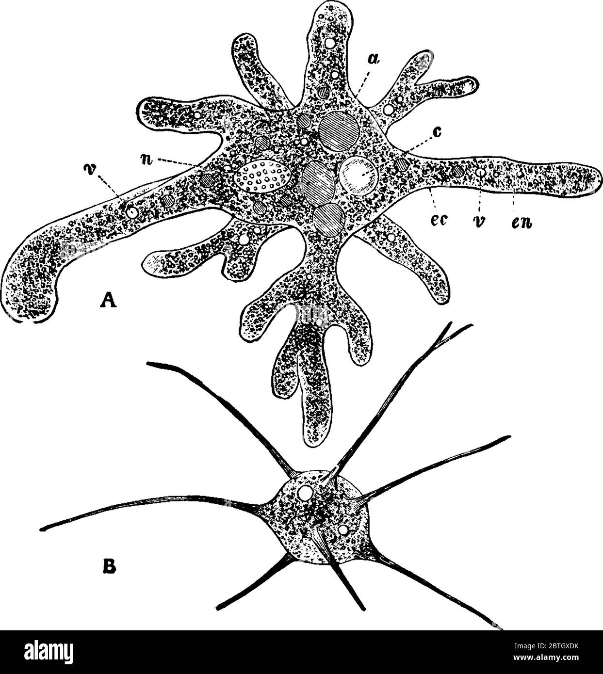
Electron Micrographs of Cell Organelles | Zoology The Electron Micrograph of Nucleus: This is an electron micrograph of nucleus. (Fig. 17 & 18): (1) Nucleus was discovered by Brown (1831). (2) It is a characteristic entity of almost all eukaryotic cells except mammalian RBCs. (3) The nucleus is generally one but may also be two, four or many.

Cellular Biology and microscopy - ppt download
Labeled Diagram Of Cell Membrane : Electron Micrograph Electron Micrograph from In other words, a diagram of the membrane (like the one below) is just a snapshot of a dynamic process in which phospholipids and proteins are continually . Some of the major parts of the plasma membrane are : How do we know what we know about cells? 1)cell membrane 2)vacuole 3)nucleus 4)endoplasmic reticulum 5)mitochondria 6)golgi body.

A Level Biology 3.2.1.1 Structure of Eukaryotic Cells ...
Electron micrographs of SPIO-labeled MSCs. A, Cell nucleus (N) and ... Download scientific diagram | Electron micrographs of SPIO-labeled MSCs. A, Cell nucleus (N) and endosomal vesicles containing SPIO particles. (Original magnification, ϫ 6600.) B, Iron-loaded ...
What is a diagram of a plant and animal cell under an ...
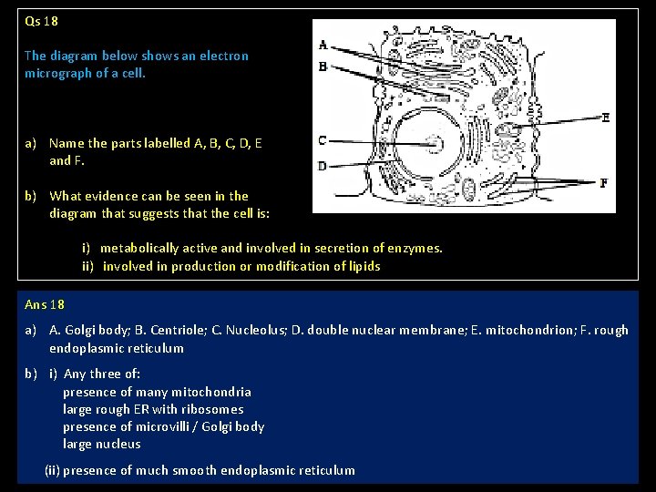
Animal Cell Electron Microscope Labelled - Q14 Draw a large diagram of ... Electron microscopes use accelerated electron beams (as opposed to visible light in a light microscope) to create images of magnification as here is an electron micrograph of an animal cell with the labels superimposed: (i) name the parts labelled as 1 to 10.

1.1 Cell structure | Cells as the basic units of life | Siyavula
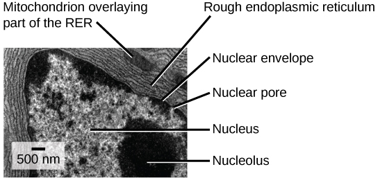
Solved Please label the electron micrograph to assess your | Chegg.com Science; Biology; Biology questions and answers; Please label the electron micrograph to assess your knowledge of the structure and function of a cell's nucleus nuclear pore endoplasma reticulum chromatin nucleolus nuclear envelope
Typical nucleus hi-res stock photography and images - Alamy
Solved Label the transmission electron micrograph of the - Chegg Label the transmission electron micrograph of the cell. 0 Nucleus rences Mitochondrion Heterochromatin Peroxisome Vesicle ULAR bumit Click and drag each label into the correct category to indicate whether it pertains to the cytoplasm or the plasma membrane.
Electron Micrographs
animal cell electron micrograph labelling Diagram | Quizlet Golgi apparatus. ... mitochondria. ... plasma membrane. ... Upgrade to remove ads. Only $2.99/month.
The Nucleus and Cytoplasm | Anatomy and Physiology | | Course ...
plant cell label electron micrograph Diagram | Quizlet Start studying plant cell label electron micrograph. Learn vocabulary, terms, and more with flashcards, games, and other study tools.
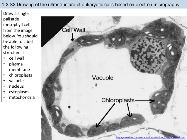
1.2 Ultrastructure of Cells - Welcome to The Frog Pad!
Cell Nucleus - function, structure, and under a microscope Summary. The nucleus is a double-layer membrane organelle. It consists of the nuclear envelope, DNA (chromatin), nucleolus, nucleoplasm, and the nuclear matrix. The main function of the nucleus is to control cell activities and carry genetic information to pass to the next generation. A eukaryotic cell typically has only one nucleus.
Ribosomes 22 nm Small organelles often attached to
DP Topic 1.1 / 1.2 | Biology - Quizizz
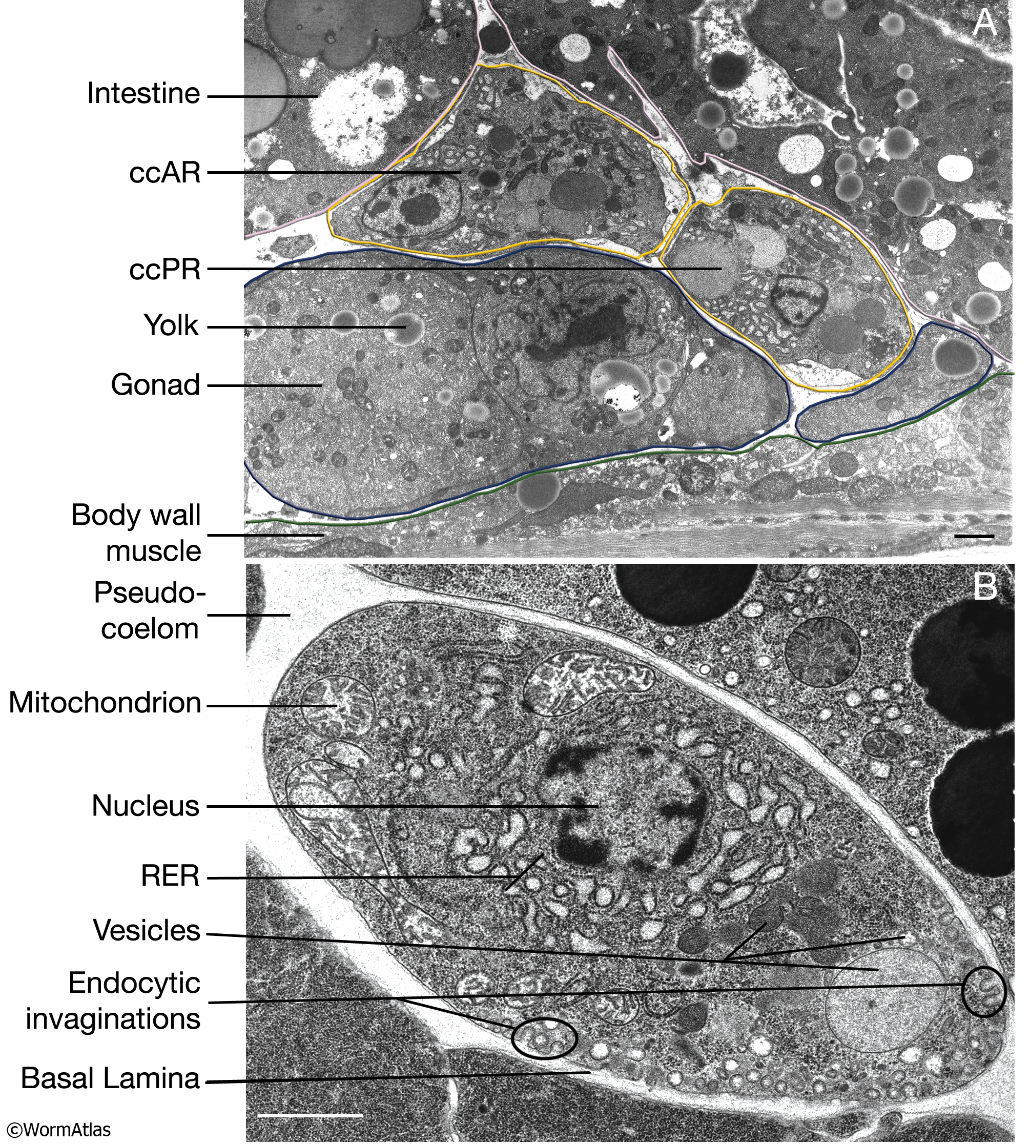
CcFIG 5 Legend

Transmission Electron micrograph section of control liver ...
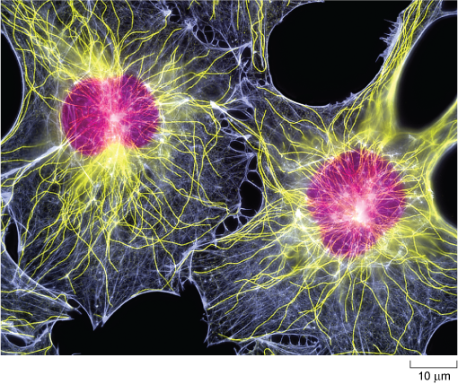
Electron Micrograph of Actin and Intermediate Filaments In ...
The Cell: The Histology Guide
Biology, The Cell, Cell Structure, The Endomembrane System ...

Electron micrographs of SPIO-labeled MSCs. A, Cell nucleus (N ...
Electron Micrographs

Electron Micrograph of Plasma Cells In Connective Tissue

AICE Biology Chapter 1: Animal Cell Electron Micrograph ...
Nucleus | Celebrate Cytochemistry | Gwen V. Childs, Ph.D.

1.1 Cell structure | Cells as the basic units of life | Siyavula
Nucleus: Definition, Structure, Functions

Normal Human Lymph Node Cells: An Electron Microscopic Study ...
Electron Micrographs
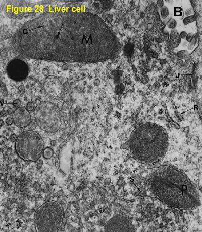
2.3.3 Identify structures from electron micrographs of liver ...
animal cell electron micrograph labelling Diagram | Quizlet

Electron micrograph of a rat hepatocyte from the control ...
The Cell: The Histology Guide

Cell nucleus - Wikipedia
A tour of the cell: View as single page

PDF) IB Questionbank Test | Ankit Mistry - Academia.edu

Electron micrograph of a rat hepatocyte from the control ...

Transmission electron micrograph of an animal cell - Stock ...
IB Questionbank

An electron micrograph of a barley nucleus including a ...
Electron Micrographs

Transmission electron micrograph of another melanocytes in ...

exocrine cell of pancreas electron micrograph labelling ...
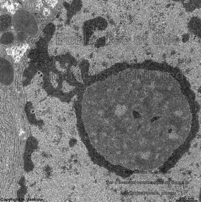
nucleolus Dr.Jastrow's electron microscopic atlas
Post a Comment for "45 nucleus electron micrograph labelled"