41 label the major bones of the skeleton
Samacheer Kalvi 6th Science Term 2 Solutions Chapter 6 Pdf Answer:Rib cage consists of 12 pairs of flat and curved rib bones. Delicate organs like heart and the lungs are well protected by this 2.) List out the functions of the human Skeleton. Answer:The major functions of the human Skeleton are: A framework is provided to the body by bones. Bones and muscles together help in movements like running ... Bone: Histology, constituents and types | Kenhub The so-called flat bones of the body such as calvaria, mandible, maxilla, etc. and long bones such as those of the limbs, are formed by two different processes. The former originates by way of intramembranous ossification, while the latter undergoes endochondral ossification.
Detailed Human Skeleton Bones Diagram - 16 images - label skeleton sjl ... [Detailed Human Skeleton Bones Diagram] - 16 images - pdhpe11 major skeletal bones, skeleton back bones diagram skeleton anatomy system human body, labeled skeleton back view of male skeleton, bones and muscles homework help skeleton and muscular system for,

Label the major bones of the skeleton
BIO 168 - Anatomy & Physiology I - Acalog ACMS™ Anatomy & Physiology I covers the structure and function of the human body at the chemical, cellular, tissue, organ, and organ system levels. Coverage includes the integumentary, skeletal, muscular, and nervous systems. Lecture and lab must be taken concurrently. List in order, from simplest to most complex, the levels of structural organization. List of skeletal muscles of the human body - Wikipedia The terms "artery" and "nerve" are both used when these structures are mentioned. Contents 1 Head 1.1 Forehead/eyelid 1.2 Extraocular muscles 1.3 Ear 1.4 Nose 1.5 Mouth 1.6 Mastication 1.7 Tongue 1.7.1 Extrinsic muscle 1.7.2 Intrinsic 1.8 Soft palate 1.9 Pharynx 1.10 Larynx 2 Neck 2.1 Clavicular 2.2 Suprahyoid 2.3 Infrahyoid 2.4 Neck 2.4.1 Anterior Skull: Anatomy, structure, bones, quizzes | Kenhub The facial skeleton is referred to as all skull bones anteroinferior to the cranial cavity. Prominent representatives are the maxilla (upper jaw) and the mandible (lower jaw). The orbita and the nasal cavity are formed by the zygomatic, nasal, palatine, lacrimal bones, the vomer and the inferior nasal concha (lower turbinate).
Label the major bones of the skeleton. BI 150 - Anatomy and Physiology I - Acalog ACMS™ define epidermis, dermis and hypodermis, listing the major layers of each and describe the function of each layer; and; discuss structures associated with epidermis, dermis and hypodermis. OSSEOUS TISSUE AND SKELETAL SYSTEM. state the functions of bones; label and study gross anatomy of a long bone; escribe the histology of Haversian system; Skeletal Muscle: Structure and Function | Exercise Physiology: Theory ... Skeletal muscles are attached to bones by connective tissue called tendons. One end of the muscle is attached to a bone that does not move (origin), and the opposite end is fixed to a bone (insertion) that is moved during muscular contraction. A variety of different movements are possible, depending on the type of joint and muscles involved. music coloring pages for adults - Unexploded Webcast Photographic Exhibit Human adults have 206 bones in their body - learn the names of the major bones. Fete De La Musique Music Coloring Music Doodle Love Coloring Pages Human Skeleton - Unlabeled You can label the major bones in this human skeleton printout. Premium Coloring Pages For Download Music Coloring Black And White Doodle Coloring Books Microscopic Anatomy Of Skeletal Muscle Worksheet The most commonly used at school since being in preschool, elementary, and the next level is the worksheet. Microscopic anatomy of skeletal muscle worksheet answers. Refer to the diagram and check your understanding of the breathing system by labeling each part and giving its functions in the box corresponding to the part.
Appendicular Skeleton: Bones List, Diagram & More - Embibe Ans: Appendicular skeleton-The appendicular skeleton includes all the bones that form the upper and lower limbs, the pelvic girdle, and the pectoral girdle. It consists of \(126\) bones. Axial skeleton-The axial skeleton is the part of the skeleton that consists of the bones of the skull, the vertebral column, and the thoracic cage. It ... Anatomy of the Human Shoulder Joint - Verywell Health Humerus (arm bone) Scapula (shoulder blade) Clavicle (collarbone) The scapula has one part that forms a socket for the ball-and-socket shoulder joint; this is called the glenoid. The glenoid is covered with smooth cartilage. The rounded top of the arm bone (humerus) contacts the shoulder blade at the glenohumeral joint. Art-Labeling Activity: Parts Of The Humerus - ARTDCA Components and divisions of the pelvis. This division of the skeleton forms the longitudinal axis of the body and protects Source: en.wikipedia.org The posterior muscles include the trapezius, rhomboid major, and rhomboid minor. When the rhomboids are contracted, your scapula moves medially, which can pull the shoulder and upper limb posteriorly. Functions of Human Skeletal System | Just-Health.net Cartilages are thick and rubbery tissues that are found in joints, the ears, the nose, and the ribs. Ligaments are tough, fibrous tissues that connect one bone to another bone, while tendons are soft tissues that connect muscles to bones. Aside from providing the body framework, there are other functions of the skeleton.
Stegosaurus - Wikipedia Stegosaurus (/ ˌ s t ɛ ɡ ə ˈ s ɔːr ə s /; lit. 'roof-lizard') is a genus of herbivorous, four-legged, armored dinosaur from the Late Jurassic, characterized by the distinctive kite-shaped upright plates along their backs and spikes on their tails. Fossils of the genus have been found in the western United States and in Portugal, where they are found in Kimmeridgian- to early Tithonian ... exercise 7 review sheet art-labeling activity 1 Exercise 38 Review Sheet Art-labeling Activity 1 1 of 2 cystic duct. Histology of Nervous Tissue. The Appendicular Skeleton Lab. Exercise 9 Review Sheet Art-labeling Activity 1 2 of 3 Drag the labels onto the diagram to identify the bones and markings of the skull. The skull the ver-tebral column and the thoracic cage. Axial Skeleton Anatomy: Diagram, Definition, Functions - Embibe The human skeleton is made up of 80 bones and is divided into six sections: the skull (22 bones), middle ear ossicles, hyoid bone, rib cage, sternum, and spinal column. The axial and appendicular skeletons combine to produce the entire skeleton. the arthropod skeleton is composed of - utekarlaxman.com the arthropod skeleton is composed of. the arthropod skeleton is composed of. June 12, 2022; liuna pension calculator; living in winters, california ...

Skeleton Anterior View - Medical Art Library
Integrating skeletal anchorage into fixed and aligner biomechanics The first temporary skeletal anchorage devices (TSADs) were retromolar osseointegrated implants to correct an acquired intermaxillary malocclusion caused by early loss of a lower first molar (L6) (Fig. 1, Fig. 2) [1,2].Small nonintegrated inter-radicular (I-R) miniscrews [3,4] were more popular TSADs that were less expensive and easier to use but not as versatile or reliable (Fig. 1B).

31 Label The Skeleton Worksheet Answers - Labels Database 2020
Dog Skeletal Anatomy - Sheridan College Scapula. Medial View of Scapula. Clincal Aspect. The scapula is a flat triangular bone at the top of the shoulder; more commonly known as the shoulder blade. It consists of 2 surfaces (medial and lateral), 3 borders (cranial, caudal and dorsal) and 3 angles (craniodorsal, caudodorsal and ventral angle). The pig and horse do not have an acromion.
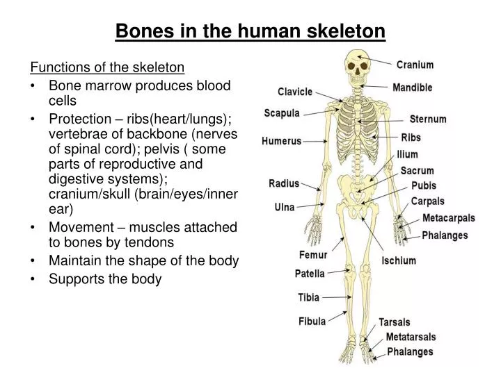
PPT - Bones in the human skeleton PowerPoint Presentation - ID:763141
Organs of Skeletal System and Their Functions - New Health Advisor The bones of human skeletal system are classified in to four distinct groups on the basis of size and shape. They are long bones, short bones, flat bones and irregular bones. The bones of arms and legs are long in length as compared to their width. Thus these are categorized under long bones.
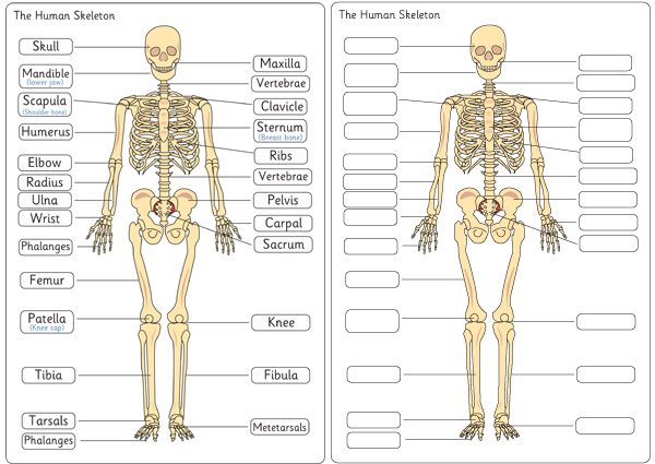
Early Learning Resources Human Skeleton Diagram Labelling Sheets
Antenatal Care Module: 6. Anatomy of the Female Pelvis and Fetal Skull 6.1.1 Ilium. Ilium is pronounced 'ill ee umm' and iliac is 'ill ee ack'. The major portion of the pelvis is composed of two bones, each called the ilium — one on either side of the backbone (or spinal column) and curving towards the front of the body. When you place your hand on either hip, your hand rests on the iliac crest, which is the upper border of the ilium on that side.

Bones of the Body
25 Major Organs of the body - Study Read Bone marrow. This is tissue, not an organ, and is present inside the large bones. It exists as yellow and red bone marrow. As a person ages, the red portion of it turns yellow. This bone marrow is essential in producing red blood cells, white blood cells, and even thrombocytes. Interstitium
Skeletal System Study Guide
What's Inside Your Bones? - Lesson - TeachEngineering The human body has 20 main bones: cranium, mandible, clavicle, scapula, vertebrae, sternum, ribs, humerus, radius, ulna, pelvis, femur, patella, fibula, tibia, carpus (carpal bones), metacarpus (metacarpal bones), tarsus (tarsal bones), metatarsus (metatarsal bones) and phalanges.

8 FREE ESL bones worksheets
What Makes Our Bones Strong? - Activity - TeachEngineering Make a chart with the written observations about what your group thinks makes bones strong. Place the chicken bone in the beaker. Cover the bone with vinegar. Cover the beaker with plastic and secure it with a rubber band. Let the bones sit four days in the liquid solution. During this time, write all observations on a chart.
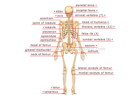
HUMAN BEING :: ANATOMY :: SKELETON :: POSTERIOR VIEW image - Visual ...
bone | Definition, Anatomy, & Composition | Britannica The two principal components of this material, collagen and calcium phosphate, distinguish bone from such other hard tissues as chitin, enamel, and shell. Bone tissue makes up the individual bones of the human skeletal system and the skeletons of other vertebrates.
Post a Comment for "41 label the major bones of the skeleton"