39 correctly label the following internal anatomy of the heart
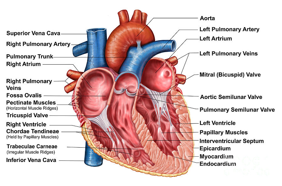
Solved Correctly label the following internal anatomy of the - Chegg Question: Correctly label the following internal anatomy of the heart. Left pulmonary veins Left ventricle Myocardium Endocardium Pulmonary trunk Interventricular septum Left atrium Epicardium Papillary muscle Left pulmonary artery Reset Zoom This problem has been solved! See the answer Show transcribed image text Expert Answer 100% (5 ratings) Solved Correctly label the following parts of the internal - Chegg Answer. Make following changes: In place of right atrium, write left atrium. …. View the full answer. Transcribed image text: Correctly label the following parts of the internal anatomy of the heart. Place your cursor over the boxes for more information papillary muscles bicuspid valve right atrium septum pulmonary semilunar valve eft atrium ...
Chapter 19: The Heart Flashcards - Quizlet The Heart •Circulatory system -heart, blood vessels & blood •Cardiovascular system -heart, arteries, veins and capillaries -2 major divisions •Pulmonary circuit - right side of heart -right heart—lungs—left heart -carries blood to lungs for gas exchange •Systemic circuit - left side of heart -left heart—body—right heart

Correctly label the following internal anatomy of the heart
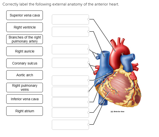
Correctly Label The Following External Anatomy Of The Anterior Heart ... Correctly Label The Following Internal Anatomy Of The Heart When you study the heart, you will find many examples of different chambers and arteries. Typically, there are four major arteries that branch out from the heart. These arteries carry oxygen-rich blood away from the heart. Correctly Label The Following Internal Anatomy Of The Heart When you study the anatomy of the heart, you will see that it has three main anatomical features. Among them are the aorta, the vena cava, and the pulmonary veins. The heart is made of tissue. It needs nutrients and oxygen. The chambers of the heart are filled with blood. However, the heart does not receive nourishment from the blood. Heart Anatomy: Labeled Diagram, Structures, Blood Flow ... - EZmed Image: Use the 2x2 table to label the 4 chambers of the heart, including the right atrium, right ventricle, left atrium, and left ventricle. Tricuspid Valve and Mitral Valve Now that we have a good understanding of the 4 chambers of the heart, let's move on to the 4 main valves.
Correctly label the following internal anatomy of the heart. Label the structures indicated on this anterior view of the heart model. Step-by-step explanation. The structures seen in the anterior view of the heart includes the superior vena cava, which transports deoxygenated blood from the head to the right atrium. The deoxygenated blood is then conveyed to the right ventricle, then to the lungs via the pulmonary trunk. The blood gets oxygen via the capillaries in the ... Human Heart (Anatomy): Diagram, Function, Chambers, Location in ... - WebMD The heart is a muscular organ about the size of a fist, located just behind and slightly left of the breastbone. The heart pumps blood through the network of arteries and veins called the ... Answered: Correctly label the following veins of… | bartleby Correctly label the following veins of the thorax. Hemiazygos v. Internal jugular v. Subclavian v. Posterior intercostal veins SubclaviarM Brachiocephalic V. Supreme intercostal v. Azygos v. Internal jugular v. Diagrams, quizzes and worksheets of the heart - Kenhub Worksheet showing unlabelled heart diagrams. Using our unlabeled heart diagrams, you can challenge yourself to identify the individual parts of the heart as indicated by the arrows and fill-in-the-blank spaces. This exercise will help you to identify your weak spots, so you'll know which heart structures you need to spend more time studying ...
AHCDW15Notes17.pdf - Course Hero Adjust credit for all students. Correctly label the following internal anatomy of the heart. Explanation: Blood that has been through the systemic circuit returns by way of the superior and inferior venae cavae to the right atrium. It flows directly from the right atrium, through the right AV valve, into the right ventricle. Label the heart - Science Learning Hub In this interactive, you can label parts of the human heart. Drag and drop the text labels onto the boxes next to the diagram. Selecting or hovering over a box will highlight each area in the diagram. Right ventricle Right atrium Left atrium Pulmonary artery Left ventricle Pulmonary vein Semilunar valve Vena cava Aorta Download Exercise Tweet Layers of the heart: Epicardium, myocardium, endocardium - Kenhub The myocardium is functionally the main constituent of the heart and the thickest layer of all three heart layers. It is a muscle layer that enables heart contractions. Histologically, the myocardium is comprised of cardiomyocytes.Cardiomyocytes have a single nucleus in the center of the cell, which helps to distinguish them from skeletal muscle cells that have multiple nuclei dispersed in the ... Chapter 20-Cardiovascular System Flashcards - Quizlet Correctly label the following internal anatomy of the heart. b Place the labels in order denoting the flow of oxygenated blood through the heart beginning with the vessels that bring blood back to the heart from the lungs. Correctly label the following coronary blood vessels of the heart.
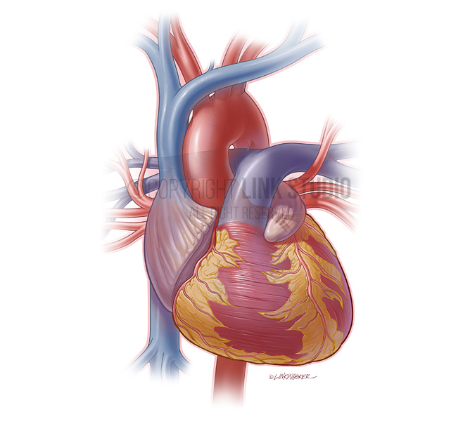
Correctly label the following external anatomy of the anterior heart. Correctly label the following external anatomy of the anterior heart. Image transcription text Ligamentum arteriosum Left pulmonary artery Left pulmonary veins Pulmonary trunk Left ventricle Ascending aorta Anterior interventricular artery Left auricle of left atrium... Show more Biology Science Anatomy Answer & Explanation Ch. 19 Circulatory System- heart Flashcards - Quizlet Correctly label the following internal anatomy of the heart. Drag each label to the location of each structure described. Explanation The heart functions to first pump deoxygenated blood returning from the body to the lungs in order to release carbon dioxide and reoxygenate the blood. Solved Correctly label the following internal anatomy of the | Chegg.com Correctly label the following internal anatomy of the heart Right atrium Interventricular septum Right ventricle Right AV (tricuspid) valve Tendinous cords Papillary muscles Fossa ovalis Right atrium Pectinate muscles Right AV (tricuspid) valve The Heart - Science Quiz - GeoGuessr The Heart - Science Quiz: Day after day, your heart beats about 100,000 times, pumping 2,000 gallons of blood through 60,000 miles of blood vessels. If one of your organs is working that hard, it makes sense to learn about how it functions! This science quiz game will help you identify the parts of the human heart with ease. Blood comes in through veins and exists via arteries—to control the ...

Electrocardiogram (ECG, EKG) Causes, Symptoms, Treatment - Basic ...
The Anatomy of the Heart, Its Structures, and Functions The heart is the organ that helps supply blood and oxygen to all parts of the body. It is divided by a partition (or septum) into two halves. The halves are, in turn, divided into four chambers. The heart is situated within the chest cavity and surrounded by a fluid-filled sac called the pericardium. This amazing muscle produces electrical ...

Heart Anatomy: chambers, valves and vessels : Anatomy & Physiology
Quiz 4 - Quiz4 1. Award: 0 out of 1.00 point Classify the following ... Award: 1 out of 1.00 point Correctly label the following parts of the internal anatomy of the heart. References Labeling Section: 05.03 3. Award: 1 out of 1.00 point Using the image as a guide, sequence the following descriptions of the flow of blood through the human heart.

Heart Anatomy External
Chapter 11 Cardiovascular System Answers Start studying the Chapter 20-Cardiovascular System flashcards containing study terms like Correctly label the following internal anatomy of the heart., Correctly label the following internal anatomy of the heart. b, Place the labels in order denoting the flow of oxygenated blood through the heart …
Anatomy Of Heart Interior, Frontal Digital Art by Stocktrek Images
Heart Labeling Quiz: How Much You Know About Heart Labeling? Here is a Heart labeling quiz for you. The human heart is a vital organ for every human. The more healthy your heart is, the longer the chances you have of surviving, so you better take care of it. Take the following quiz to know how much you know about your heart. Questions and Answers. 1.
/heart_interior-570555cf3df78c7d9e908901.jpg)
Heart Wall: Epicardium, Myocardium, and Endocardium
Cardiovascular System Heart Chapter 20 - hex.arista.com Start studying the Chapter 20-Cardiovascular System flashcards containing study terms like Correctly label the following internal anatomy of the heart., Correctly label the following internal anatomy of the heart. b, Place the labels in order denoting the flow of oxygenated blood through the heart beginning with

Februari 2011
Solved Correctly label the following parts of the internal - Chegg Correctly label the following parts of the internal anatomy of the heart. Right pulmonary veins Aorta Left pulmonary veins Pulmonary semilunar Left ventricle Right ventricle Bicuspid valve Tricuspid valve Right pulmonary artery Septum Pulmonary trunk Right atrium Left pulmonary artery Inferior vena cava Left atrium Superior vena cava Reset Zoom

36 Label Heart Parts - Labels 2021
Chapter 19 Homework Flashcards - Quizlet correctly label the following internal anatomy of the heart Which of the following carry oxygen-poor blood. a. Pulmonary veins and vena cavae. b. Aorta and pulmonary veins. c. Aorta and vena cavae. d. Venae cavae and pulmonary arteries. e. Pulmonary veins and pulmonary arteries. venae cavae and pulmonary arteries
Heart Anatomy External
Anatomy & Physiology: The Unity of Form and Function - Quizlet Correctly label the following coronary blood vessels of the heart. Correctly label the pathway for the cardiac conduction system. Drag each label to the location of each structure described. Vessel carrying oxygenated blood to the myocardium. Artery carrying deoxygenated blood. First vessel of systemic circuit. Veins carrying oxygenated blood.
31 Label This Anterior View Of The Human Heart - Labels Database 2020
AHCDW15Notes14.pdf - Course Hero Correctly label the following internal anatomy of the heart. Explanation: The anterior and posterior interventricular sulci overlie an internal wall, the interventricular septum, that divides the right ventricle from the left. The coronary sulcus and two interventricular sulci harbor the largest of the coronary blood vessels.
Post a Comment for "39 correctly label the following internal anatomy of the heart"